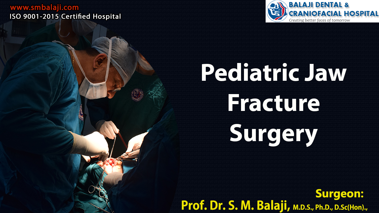
The patient is a 4-year-old boy from Kayalpatnam in Tamil Nadu, India. He had been playing cricket and had run across the road to collect the ball. A motorcycle had collided with him and he had landed hard with his jaw hitting the asphalt. There was a laceration at the symphyseal region and extreme pain. The motorcyclist had left the scene immediately after the accident.
His concerned parents had immediately taken him to a nearby hospital for treatment. The laceration was debrided and dressing was done. An x-ray revealed a fracture in the anterior region of the mandible. Tetanus toxoid had also been administered to the patient at the hospital. He was advised to undergo fracture fixation with plates and screws.
Patients advised against usage of plates and screws for child
However, the parents were advised against this by a family friend who is an orthopedic surgeon. The orthopedic surgeon then referred the patient and his parents to our hospital for management of his fracture. The bones of children are unlike adult bones. An adult bone breaks easier while a child’s bone bends.
Fractures in children heal faster. Care should be taken to not disrupt the growth plates. Closed reduction with active range of motion is the ideal treatment for simple childhood fractures. Our hospital follows all the surgical protocols laid out for pediatric fracture surgery by the American Association of Oral and Maxillofacial Surgeons.
Our hospital is a premier center for pediatric oral and maxillofacial surgery in India. We are a central referral center for cleft lip and cleft palate surgery in Southeast Asia. Pediatric fracture surgery is routinely performed in our hospital. All the latest radiographic imaging modalities are available in house for easy diagnosis and treatment planning of cases.
Initial presentation at our hospital
Dr SM Balaji, Pediatric Fracture Surgeon, examined the patient and ordered comprehensive diagnostic testing for the patient. The patient was unable to open his mouth. The parents stated that he was not able to chew food and that his appetite had gone down considerably following the accident.
Imaging studies obtained included an OPG as well as a 3D CT scan. These revealed a symphysis fracture and a displaced left condylar fracture.
Surgical treatment planning for the patient
The patient’s parents were advised that the symphysis fracture was best addressed through a cap splint. It was also explained that fixation of the condylar fracture would be unnecessary as the occlusion would be maintained by the cap splint. This would allow for total union of his condylar fracture.
It was also advised that he would need to stay on a liquid diet for about 2-3 weeks followed by a semi-solid diet. The patient was scheduled for pediatric jaw fracture surgery for his pediatric condylar fracture correction.
Successful surgical correction of the fracture
Under general anesthesia, an impression was taken of the mandibular arch and a cap splint fabricated. The cap splint was then placed on the lower jaw and wired using circum mandibular wiring. Chin laceration was also sutured using resorbable and nonresorbable sutures. The fracture site was thus reduced using cap splint, thereby limiting movement of the jaw and enabling bone healing.
Parents of patient express complete satisfaction
The surgery was successful with no complications. Parents were very satisfied with the outcome of the surgery. After a period of about one month, the wires were removed under general anesthesia after the fracture site showed considerable healing.
The patient was able to open and close his mouth again with no pain or discomfort. Parents were very thankful as they had been initially worried about the possible long term complications that could affect the boy’s quality of life.
Patient develops a deformity of the left side of the face
The patient is a 44-year-old patient from Bilaspur in Chattisgarh, India. He was fine until approximately two years ago. It was then that he began noticing a swelling develop on the left side of his lower jaw. Alarmed by this, he had presented at a local maxillofacial hospital for diagnosis and management.
A detailed examination had been performed followed by comprehensive imaging studies and a biopsy. The patient had been diagnosed with an odontogenic keratocyst. He had been advised plastic and reconstructive surgery for management of his condition. Of note, the patient himself is a medical doctor.
Characteristics of an Odontogenic Keratocyst
An odontogenic keratocyst is a rare, benign but locally aggressive developmental cyst. It most often affects the posterior mandible and most commonly presents in the third decade of life. Odontogenic keratocysts make up around 19% of all jaw cysts. Radiographically, most are unilocular in presentation when present at the periapex. They can be mistaken for a radicular or lateral periodontal cyst. Removing the cyst completely would entail removal of the surrounding tissues.
There are many types of cysts that occur in living organisms. A dentigerous cyst occurs in relation to an impacted tooth. Other cysts include sebaceous cysts, epidermoid cysts, pilar cysts and ovarian cysts. Recurrence rates vary with the kind of cysts. Pilar cysts are associated with infected hair follicles. Odontogenic keratocysts sometimes occur in conjunction with nevoid basal cell carcinoma syndrome.
Initial surgical intervention for his odontogenic keratocyst
The patient had undergone a hemimandibulectomy around 3 years ago for removal of his odontogenic keratocyst. His mandible was then reconstructed using titanium plates and screws. This had resulted in a gross deformity of the left side of his face. The screws of the plate also showed evidence of loosening. He also experienced extreme difficulty with chewing and speech. This had left him very depressed and he sought the help of his friends in finding a solution to his problem.
His friends searched far and wide for the best hospital to treat him. Their enquiries finally convinced them that our hospital was the right choice for him. They then informed the patient that his problem would be best addressed at Balaji Dental and Craniofacial Hospital in Chennai. The patient and his wife then made an appointment to meet Dr SM Balaji, facial deformity surgeon.
Initial presentation at our hospital for management and treatment
Dr SM Balaji, cyst removal surgeon, examined the patient and studied his old case records in detail. He then ordered comprehensive imaging studies including a 3D CT scan for the patient. The patient had a gross deformity of the face with a left-sided asymmetry. There was also a slight deviation of the mandible to the left side.
Imaging studies revealed the presence of the titanium reconstruction plate fixed to the left condyle with loosened screws. There was no recurrence of the odontogenic keratocyst at the site.
Formulation of treatment plan for the patient
The patient expressed that he was not interested in undergoing microvascular reconstructive surgery. Hence it was explained to the patient that reconstruction with rib graft surgery would offer the best results. This would involve harvesting a rib graft from the patient at the time of surgery.
It was also explained that dental implants would be placed once the rib grafts are consolidated. This would ensure return to complete normal function for the patient. The patient was in agreement with the treatment plan and consented to surgery.
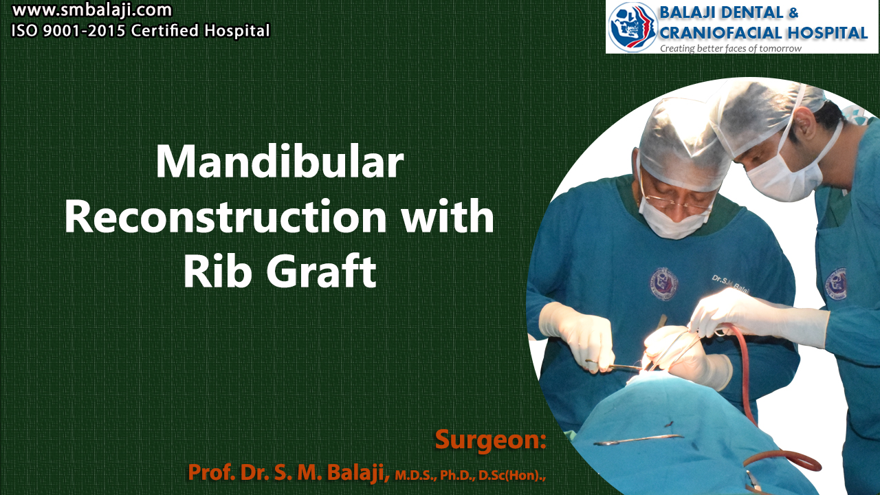
Successful surgical correction of the patient’s facial deformity
Under general anesthesia, a right inframammary was incision placed and dissection was carried down to the ribs. A costochondral rib graft was then harvested to be used for the mandibular graft surgery. A Valsalva maneuver was then performed to ensure that there was no perforation into the thoracic cavity. The incision was then closed in layers using sutures.
Attention was then turned to the reconstruction of the mandible. A midcrestal incision was made in the left mandible followed by elevation of a mucoperiosteal flap. Mandibular reconstruction was then performed using the rib graft along with the same mandibular reconstruction plate. Hemostasis was achieved followed by closure of the incision with sutures.
Complete patient satisfaction with the results of the surgery
The patient was very happy with the outcome of the surgery. There were no scars present as all the incisions had been made intraorally. Facial asymmetry had been achieved with the reconstruction of the mandible. His facial appearance was back to baseline following surgery. It was explained that dental implants would be placed on the bone graft after consolidation of the grafts. This would be followed by placement of crowns after complete osseointegration of the implants.
The patient expressed complete understanding of the future treatments and would return in 3-4 months for placement of dental implants.
Patient with difficulty breathing and snoring problems
The patient is a 25-year-old male from Cuttack in Odisha, India. He had always had difficulty with breathing since childhood. A whistling sound would sometimes emanate from his nose while breathing. This is one of the classic symptoms of a deviated nasal septum. The septum is the cartilage that divides the nostrils. Surgery is usually required to correct a deviated septum.
Difficulties at school because of poor sleep at night
He also has always been plagued by snoring since childhood. The quality of sleep had always been poor and he had chronic daytime sleepiness. His studies had been affected because of daytime sleepiness with complaints from teachers.
He was also not satisfied by the appearance of his nose, which he felt had a depressed base. The nose is essentially made of bone and cartilage. His nasal appearance always made him feel self conscious and he tended to avoid social gatherings because of this. He had always desired to have a prominent and elevated nose.
Deciding to get this corrected, he had visited a rhinoplasty surgeon in his hometown. Upon examination, the patient was found to have a deviated nasal septum. He had undergone a nose correction surgery at that time, but was not satisfied with the results.
The patient had then approached another plastic surgeon locally. He had explained to the patient that he still had the nasal deformity and that this needed to be surgically corrected at a specialty center. He had then referred the patient to our hospital for correction of his nasal deformity. The surgical cost in India is negligible compared to the Western countries.
Our hospital is a premier center for cosmetic rhinoplasty in India. Thousands of patients have been rehabilitated in our hospital from their breathing difficulties over a period of 25 years. The results of the surgery have always directly resulted in improvement in the quality of their lives.
Breathing difficulties arising from nasal deviation always affect the quality of sleep. Correction of the deviation results in immediate improvement in the quality of sleep.
Initial presentation and treatment planning at our hospital
Dr SM Balaji, a premier rhinoplasty surgeon in India, examined the patient and ordered for comprehensive radiological imaging studies. This revealed that the patient had a deviated nasal septum. There was also a depressed nasal dorsum with a hanging alar base. A treatment plan was formulated to correct the patient’s complaints.
A septoplasty or deviated nasal septum surgery was planned for correction of the deviated nasal septum. Augmentation of the nasal bridge would be through a costochondral rib graft. Alar base correction would also be performed at the same time.
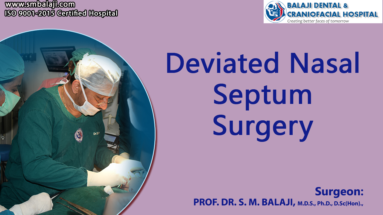
Surgical correction of the patient’s nasal deformity
Under general anesthesia, an incision was made in the right inframammary region. This was followed by dissection down to the ribs. A costochondral rib graft was then harvested. A Valsalva maneuver was then performed to ensure that there was no perforation into the thoracic cavity. The incision was then closed in layers with sutures.
Attention was then turned to correction of the nasal deformity. A transcartilaginous incision was made in the right nostril and dissection was done up to the nasal dorsum. This was followed by an intercartilaginous incision with dissection down to the medial nasal cartilage.
A septoplasty was then performed. The nasal dorsum was augmented using the costochondral rib graft. Attention was next turned to the nasal sill, which was corrected by excising a portion of nasal mucosa. Intranasal suturing was then done using resorbable sutures.
Total patient satisfaction with the results of the surgery
The cosmetic result of the surgery was immediate. He now had a more elevated and symmetrical nose. The nasal deformity had been completely corrected and the patient was extremely happy with the outcome of the surgery. He expressed that he was able to breathe well and that the quality of his sleep was drastically improved.
The patient is a 26-year-old male from Kurnool in Andhra Pradesh, India. He was born with a left-sided cleft lip and palate. Cleft lip surgery and cleft palate surgery were performed before the patient was a year old. A cleft alveolus surgery was performed at the age of 7 years. The hole in the roof of the mouth was closed. All the surgeries had been performed elsewhere.
The surgeries had left the patient with a deformed nose and a prominent hypertrophic scar in the upper lip. There was no hair growth in the region of the scar, which upset the patient as he desired to have a mustache.
The nose was flattened and asymmetrical. He had faced a considerable amount of bullying at the hands of his peers, both in school and in college. This had made him feel depressed and reclusive. The patient always desired to possess a sharp nose that was in harmony with the rest of his face. He also said that he felt he had a decreased sense of smell and breathing problems.
His parents realized that he required plastic surgery to address his problems. He and his parents approached many plastic surgeons and facial cosmetic surgeons for correction of his deformity. They were then referred to our hospital for surgical correction of his complaints. Our hospital is a renowned specialty center for the surgical correction of cleft lip, palate and alveolus deformity.
Cleft lip, palate and alveolus etiology and ramifications
Absence or incomplete fusion between the segments of the two halves of the upper lip segments in utero lead to the formation of a cleft lip, palate and alveolus. It could occur either due to environmental factors, injuries to the fetus, fetal infections or could have an underlying genetic basis to it. It is the most common birth defect found in newborns.
Scientific research is being carried out around the world to find a way to prevent the formation of cleft lip and palate. This is very distressing to both the patients once they grow old enough to become aware and to the parents.
There was a lot of stigma attached to this during the prescientific days. Better scientific understanding into the causes of cleft lip and palate has removed the stigma attached to it. Surgical correction is the only way to address this congenital facial deformity.
Initial presentation at our hospital for correction of his deformities
Dr SM Balaji, Facial Deformity Surgeon, examined the patient and obtained comprehensive imaging studies including 3D CT scan. All vitals were checked including blood pressure. Treatment planning was then made for the patient. The patient would need nose correction surgery, also known as a nose job.
It was explained to the patient and his parents that nasal bridge augmentation would be performed with a costochondral rib graft. A strut graft would then be utilized to elevate the left nostril by means of lifting up the columella. A nose essentially has both bone and cartilage in it.
Medial and lateral osteotomies would then be performed to correct the deformity along with lip revision for scar removal. The patient and his parents were in agreement with the treatment plan and consented to surgery.
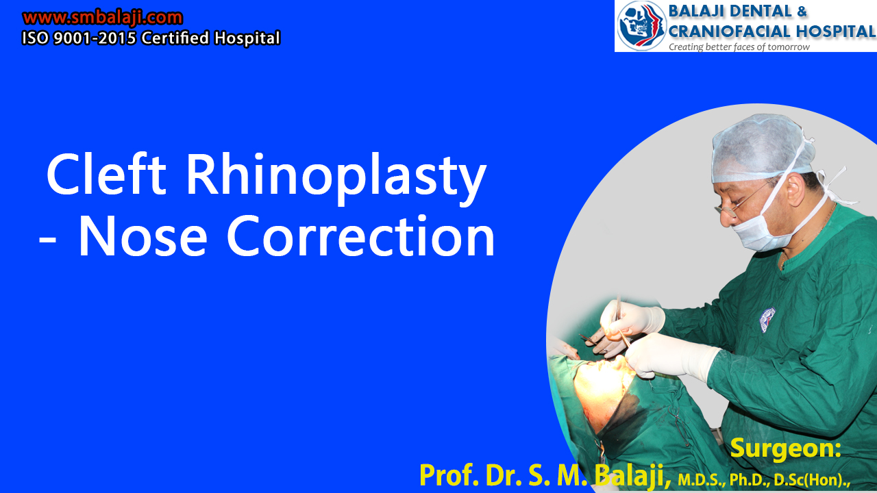
Successful surgical correction of the deformities
Under general anesthesia, an incision was made in the right inframammary region and dissection was carried down to the ribs. A costochondral rib graft was then harvested followed by a Valsalva maneuver to ensure a patent thoracic cavity. The incision was then closed in layers with sutures.
An intercartilagenous incision was then made followed by medial and lateral osteotomies to correct the deviated nasal septum. Once nasal septum surgery had been done, dissection was then done and the nasal bridge was augmented with the costochondral graft. A strut graft was then placed to elevate the left nostril.
Scar revision surgery followed with excision of scar tissue from the upper lip. Closure was then done intranasally using resorbable sutures and extraorally using non-resorbable sutures.
Complete patient satisfaction at the outcome of the surgery
The patient and his parents were very happy with the results of the surgery. The patient now had a sharp, symmetrical and prominent nose after the cleft rhinoplasty. Upper lip scar revision surgery also enhanced the esthetics of the patient’s face.
He expressed his joy to the surgeon and mentioned that this will help him to lead a life with more confidence.
Patient born with a unilateral cleft lip and palate
The patient is a 28-year-old male from Nashik in Maharashtra, India who was born with a cleft lip and palate. These are the two most common birth defects found in newborns. Speech development is also affected by these defects. Breathing problems can also be associated with them. The surgical procedures require extreme precision and dexterity for successful esthetic outcomes.
He had undergone cleft lip surgery at 3 months of age and cleft palate surgery at 9 months of age by a cleft team. The hole in the roof of the mouth had been closed. Both surgeries had been performed elsewhere.
However, the patient’s nose had become progressively deformed as he grew up. It was flat and crooked and the patient felt that it made him unattractive. This had become more and more noticeable with the passing years.
Social problems arising from the presence of the scars
There was also a very prominent scar from the cleft lip surgery. This had caused a great deal of distress to the patient and had made the patient an introvert. He rarely left the house and had few friends. There had also been instances of bullying at school and college. His lip and nose deformities played on his mind. He wanted to undergo cosmetic surgery for surgical repair of his nose and lip.
The patient had consulted cosmetic surgeons and plastic surgeons in his hometown who had examined the patient. Realizing the degree of correction required, he had referred the patient to our hospital for rhinoplasty and lip revision surgery. A rhinoplasty is commonly referred to as a nose job.
Center of excellence for cosmetic nose surgery
Our hospital is a specialty center for cosmetic rhinoplasty. All forms of nasal deformities are corrected surgically at our hospital. Bone grafts are used to address many of these procedures. Even many celebrities have undergone cosmetic nose surgery here. Subtle changes in the form of their nose even gave some of them a new lease in their career.
Initial examination and treatment planning at our hospital
Dr SM Balaji, cosmetic nose surgeon, examined the patient and and ordered comprehensive imaging studies. The patient stated that he wanted a sharp, prominent and symmetrical nose. He also stated that he was very self conscious about the residual scar from his cleft lip surgery. There was loss of mustache from the philtrum.
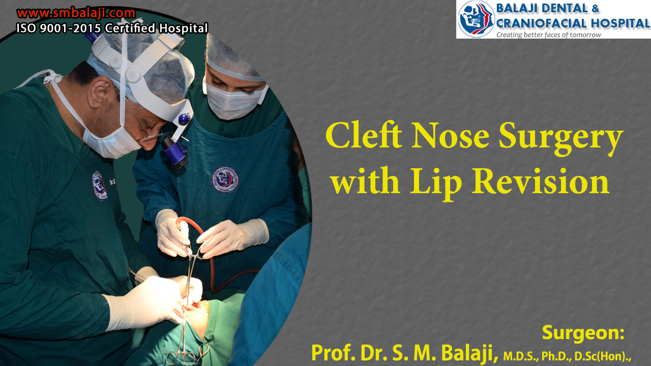
The patient also requested scar revision surgery. Lip revision followed by nose correction with costochondral rib graft was planned for the patient. Right lateral osteotomy was also planned to correct the nasal deformity. The patient was scheduled for cleft nose surgery with lip revision.
Successful surgical correction of deformed nose and lip
Under general anesthesia, the lip revision surgery was performed first with excision of the residual scar tissue. Following this, an incision was made in the right inframammary region and a costochondral rib graft was harvested.
A transcartilagenous incision was next made in the right nostril with dissection done up to the nasal dorsum. The nasal dorsum was augmented using the costochondral graft. A right lateral osteotomy was then done followed by placement of resorbable sutures intranasally.
Total patient satisfaction at the outcome of the surgery
The patient was extremely happy with the results of the surgery. His nasal asymmetry had been corrected and there was perfect harmony with the rest of the face. He no longer had a depressed or crooked nasal form. His nose was symmetrical, elevated and prominent. The scar revision was also successful.
He stated that he can now face life with confidence with the correction of his nasal and lip deformities. This would also help him be less introverted and develop an active social life.
The patient is a 41-year-old male from Chikkaballapur in Karnataka, India. He noticed the development of a swelling in his right lower jaw around 9-10 months ago. Thinking it would subside on its own, he had ignored it for a couple of months. It was the development of pain, which made him approach a dentist for consultation.
This had progressively increased in size along with development of pain and loosening of teeth. Chewing and eating became progressively difficult because of the swelling. Alarmed at this, he had presented to a local oral surgeon for diagnosis and management. Radiographs had been obtained revealing the presence of a dentigerous cyst in the region.
Seeing the complicated presentation of the cyst, the surgeon referred the patient to our hospital for management. Our hospital is a renowned center for dentigerous cyst surgery in India. We are also a premier center for mandibular reconstruction in India.
Dentigerous cyst:
A dentigerous cyst is an odontogenic cyst, which is felt to be of developmental origin. This is always associated with the crown of an unerupted or a partially erupted tooth. Mandibular third molars are the teeth most commonly associated with a dentigerous cyst. These are the most commonly impacted teeth in the mouth.
Next most commonly involved teeth are the maxillary canines. They are also rarely found involving deciduous teeth and occasionally odontomas. Orthodontic treatment is employed to bring the canines into the arch if there is no pathology present in relation to the impacted tooth.
Diagnosis is through radiographic imaging of the affected region. Dentigerous cysts are classified according to their radiological position, namely central type, lateral type and circumferential type. A dentigerous cyst is often treated by excision of the cyst along with the extraction of the associated tooth.
Initial presentation at our hospital
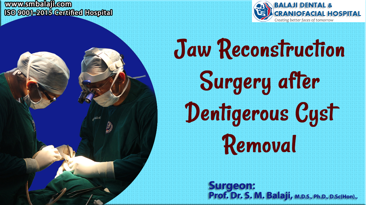
Dr SM Balaji, jaw reconstruction surgeon, examined the patient and ordered comprehensive imaging studies including a 3D CT scan. The patient had a swelling in relation to right lower molar region. Imaging studies revealed the presence of a cystic lesion extending from the first molar up to the ramus of the mandible. This confirmed the diagnosis of dentigerous cyst.
Treatment planning explained to the patient
It was explained that the cyst needed to be enucleated completely. Associated teeth would also be extracted to ensure that the cyst did not recur. This would be followed by jaw reconstruction surgery. The residual bony defect would then be reconstructed using a costochondral graft harvested from the patient.
Final step of rehabilitation of the patient would involve dental implants. This would enable the patient to regain full masticatory function of his jaws. Abutment is attached to the implant. Artificial teeth are then attached to the implants to facilitate this. Implants mimic tooth roots. Good gum tissue health is necessary for success of implants. Single implants can be placed under local anesthesia.
Dental implants would be placed after consolidation of the grafts. This would result in complete rehabilitation of the patient’s oral cavity. Dental implants need meticulous oral hygiene and dental care for optimal results in the long run. In the case of full mouth rehabilitation with implants, removable dentures can be placed over the implants.
Successful surgical enucleation of the dentigerous cyst
Under general anesthesia, a right inframammary incision was first made. This was followed by dissection down to the ribs following which a costochondral rib graft was harvested. A Valsalva maneuver was then performed to ensure that there was no perforation into the thoracic cavity. The incision was then closed in layers with sutures.
Following this, a crevicular incision was made in the mandible and a mucoperiosteal flap was elevated. The right lower first and second molars were then extracted as they contacted the cystic cavity. A transalveolar extraction of the impacted third molar was then performed followed by total cyst enucleation.
This was followed by electrocauterisation of the cystic cavity along with antibiotic flushing. The resultant bony defect was packed with rib graft. Hemostasis was check followed by closure of the incision with sutures. There was satisfactory control of postoperative bleeding at the surgical site.
Complete patient satisfaction at the surgical result
The patient expressed complete satisfaction at the surgical results. There was no external scar as all incisions were intraoral. It was explained that dental implant surgery done after consolidation of bone would result in total rehabilitation.