The patient is a 30-year-old male from Jamshedpur in Jharkhand, India. Growth of his facial bones was normal until he became a teenager. It was around that time that the right side of his lower face began to develop an asymmetry. This slowly but gradually worsened to the point where he now has gross asymmetry of his face.
Eating most foods had also become very difficult because of an increasing left posterior open bite. He also soon developed pain in his right temporomandibular joint. These two issues have been present for over 10 years now. He had also faced significant bullying during his school and college days. This has made him an introvert.
Initial surgery for correction of asymmetry in a nearby state
Desiring to get this treated, he had approached a maxillofacial surgeon in a neighboring state for consultation. A detailed examination had been performed and he had been advised surgery. He had subsequently undergone reduction of the mandibular angle and shortening of the ramus on the right side. A piece of the ramus had been removed and the ends brought together with titanium plates and screws.
The patient however was greatly disappointed by the results of the surgery. He was left with an unsightly scar on the right side of his face due to the extraoral approach adopted by the surgeon. The patient also realized that his chewing problem and pain remained the same. There was also an increase in the muscle mass on the right side of the face. There was also an increase in his facial asymmetry.
He had approached another oral and maxillofacial surgeon who had advised bone grafting from the iliac crest(hip bone graft). The patient’s mandibular deficiency caused by the first surgery was addressed through the use of this graft, but this led to compromised facial esthetics for the patient.
Referral to our hospital for treatment of his facial deformity
Feeling despondent by the turn of events, he had approached a local surgeon in his hometown. Upon examining the patient and realizing the magnitude of damage caused by the first two surgeries, the surgeon had immediately referred the patient to our hospital. Our hospital is renowned for facial asymmetry correction in India. Facial asymmetry surgery is routinely performed utilizing distraction osteogenesis surgery.
Initial presentation for treatment at our hospital
Dr SM Balaji, facial asymmetry surgeon, examined the patient and obtained a detailed oral history. He also ordered for comprehensive imaging studies including a 3D CT scan. It was clear that this was a case of idiopathic hyperplastic right mandibular ramus.
Clinical examination revealed an open bite on the left side and increased muscle mass on the right side of the face; however, OPG revealed that the right mandible was short by 10 mm. The 3D CT scan also revealed defective mandibular ramus length on the right side as well. Iatrogenic damage from the previous surgeries was also clearly visualized in the imaging studies.
Treatment planning explained in detail to the patient
Dr SM Balaji explained that correction of his asymmetry required right mandibular ramus distraction osteogenesis. A distraction of about 10 mm was planned for the patient. Subsequent rib grafting was planned for correction of the iatrogenically induced mandibular angle defect.
Distraction devices can be classified into external devices and internal devices. Oral and Maxillofacial Surgery predominantly uses internal devices. Orthopedic surgery however mainly relies on external devices. Completion of the distraction process is always followed by the consolidation phase. This is for the bone at the surgical site to be strengthened.
Successful placement of the Univector mandibular distractor
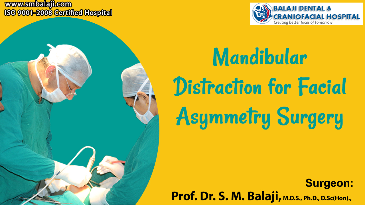
Under general anesthesia, an incision was first placed in the right mandibular posterior region. This was followed by elevation of a flap. Horizontal bone cuts were then made and the Univector mandibular ramus distractor was fixed using titanium screws. Distractor function was checked and was found to be optimal.
Following the placement of the distractor, a sulcular incision was placed in the maxilla. Le Fort 1 bone cuts were made and the maxilla was mobilized. The posterior end of the left maxilla was fixed using transosseous wires. This would enable correction of the occlusal cant. Hemostasis was achieved and closure was done using resorbable sutures.
Postsurgical distraction performed for asymmetry correction
Interarch wiring was done. A latency period of about 6-7 days was allowed for settling down of the surgical site. Following the latency period, the distractor was activated by 1 mm each day. After achieving a satisfactory increase in length of 10 mm, the distraction was stopped. Two weeks following completion of distraction, a straight plate was fixed to the left posterior maxilla to prevent further downward movement.
Total patient satisfaction with the results of the surgery
The patient was extremely satisfied with the results of the surgery. His facial asymmetry had been corrected and his entire face was in harmony. The patient’s parents were also very happy with the results and said that the patient had become noticeably happier following surgery.
They were instructed to return after a few months for removal of the distractor after bony consolidation had been demonstrated at the surgical site. The patient and his parents expressed understanding of the same.
Patient struggling with facial asymmetry deformity
The patient is a 16-year-old male from Secunderabad in Telangana, India. His parents state that he had been born with a deformity of the mouth with a right lateral facial cleft. This is commonly known as macrostomia. He had undergone surgery for correction of his microstomia during his childhood. The parents stated that the patient has always had residual scarring from that surgical procedure.
A gradually developing facial asymmetry was soon noted by the parents with the passage of time. There was underdevelopment of the right side of the face, which was becoming worse. The patient had undergone testing, which had returned with the diagnosis of hemifacial microsomia. It has now reached the point where the patient’s face demonstrated extreme asymmetry of the two sides.
The patient had become completely dejected and depressed by this progressive development of facial deformity. He had faced a tremendous amount of bullying at school, which had made things worse for him. It has now reached the stage where he is refusing to attend school or at times even leave the house to attend social gatherings.
The parents then began their quest at finding the right hospital for their son’s treatment. They had made enquiries all over the country. These enquiries had finally led them to our hospital. Our hospital is a premier center for hemifacial microsomia surgery in India. Scores of patients have been successfully rehabilitated in our hospital and are now leading normal lives.
Hemifacial microsomia and lateral facial clefts
Hemifacial microsomia is a congenital disorder that affects the development of the lower half of the face. It most commonly affects the ears, the mouth and the mandible. It usually occurs on one side of the face, but rarely involves both sides. When severe, it may result in breathing difficulties due to obstruction of the trachea, which might even require a tracheotomy.
Incidence of hemifacial microsomia is in the range of 1:3500 to 1:4500 live births. This is the second most common birth defect of the face after cleft lip and cleft palate. Lateral facial clefts arise from the failure of the maxillary and mandibular prominences to fuse at the lateral commissure. This gives rise to macrostomia.
Initial presentation and consultation at our hospital
Dr SM Balaji, facial asymmetry surgeon, examined the patient and ordered for comprehensive imaging studies. The patient had a noticeable scar on his right cheek from the lateral facial cleft correction. There was also a gross facial asymmetry on the right side. The patient also had an occlusal cant due to his mandibular deformity. A 3D CT scan revealed a deformed right mandible with a hypoplastic ramus.
It was explained that he needed to undergo mandibular ramus distraction osteogenesis surgery. This would be on the right side and would correct his facial asymmetry. A Le Fort I maxillary osteotomy was also planned for correction of the asymmetry and the occlusal cant. Facial symmetry is established when it is used for correction of asymmetrical mandible.
Clinical application of distraction osteogenesis covers the entire skeleton. It is used for limb lengthening in case of limb length discrepancy. This is very safe and the resulting bone structure is both stable and strong. Soft tissue molding also happens concurrent with the bone lengthening. Bone grafts are unnecessary for this procedure.
Successful surgical correction of hemifacial microsomia deformity
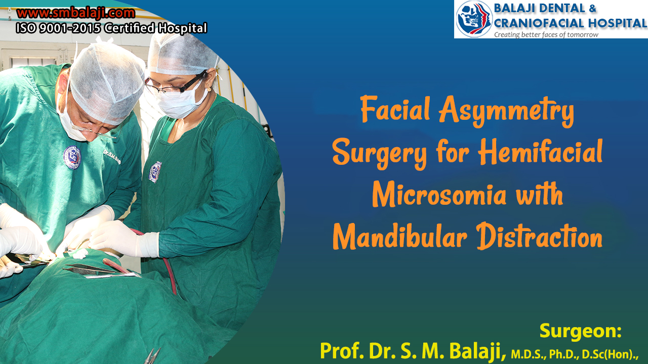
Under general anesthesia, an incision was first made in the right submandibular region. Dissection was done up to the right mandibular ramus. This was followed by horizontal bone cuts to the outer cortex following which the mandibular ramus distractor was fixed using titanium screws. The inner cortex was then separated. Extreme care was taken to protect the inferior alveolar nerve throughout the procedure.
Following this, a sulcular incision was made in the maxilla followed by a Le Fort I osteotomy. The maxilla was then mobilized. Hemostasis was achieved and closure was done using resorbable sutures. Interarch wiring was then done to stabilize the surgical site.
Postsurgical phase of the treatment
A latency period of about six to seven days was allowed after surgery for stabilization of the surgical site. Following the latency period, activation of the distractor was begun. This was by 1 mm every day for a period of 25 days to achieve a total mandibular advancement of 25 mm on the right side. Distraction was stopped after this period. A period of two more weeks were allowed before fixation of a straight plate to the left posterior maxilla to prevent further downward movement.
Successful completion and rehabilitation of the patient
After a period of about four months, radiographs were obtained to evaluate the site of distraction. This revealed complete consolidation of the bone with a reformation of a patent inferior alveolar nerve canal. The patient was extremely happy with the esthetic results of the surgery. He had a symmetrical face as well as normal occlusion with stabilization of the occlusal cant.
The patient is a 20-year-old female from Kottayam in Kerala, India who has a disproportionately large lower jaw. This problem manifested at an early age in the patient. She said that she has always had a very long lower jaw, which had gradually worsened with time. Her parents had sought a consultation with a local oral surgeon who had advised surgical correction after completion of her growth.
Thumb sucking habit results in the exact opposite of this. It causes maxillary prognathism and a class II skeletal malocclusion. It is essential that habits like thumb sucking are broken at the earliest to avoid later problems with the jaw bones and teeth. The tongue posture is itself changed by such habits.
Effects of adverse effects on future treatment needs for the patient
This leads to requiring extensive treatment to obtain normally positioned permanent dentition. Such treatments could include both surgery as well as orthodontics. Treatment can be initiated during the mixed dentition phase.
The patient is now 20 years old and had approached him again. Seeing the degree of correction needed for the patient, he had referred them to our hospital for correction of her problem. The patient did have an extreme degree of mandibular prognathism.
Her mandibular prognathism is so extreme that she is unable to close her mouth. She also has a lot of difficulty with chewing and speech. Our hospital is a premier center for corrective jaw surgery in India. Both jaw reduction surgery and jaw advancement surgery are done at our hospital. Her mandibular plane would also be normalized by this surgery.
Distraction osteogenesis is the treatment of choice for increasing jaw size. Reduction of mandibular size is through bilateral sagittal split osteotomy.
Occurrence of mandibular prognathism with possible etiology
Prognathism in humans can be due to normal variation among phenotypes. In humans, prognathism may be a malformation, the result of injury, a disease state or a hereditary condition. It is considered a disorder only when it affects mastication, speech or the ability to function normally in a social setting.
Mandibular prognathism is a protrusion of the mandible in relation to the rest of the face. Pathologic mandibular prognathism is a potentially disfiguring genetic disorder where the lower jaw outgrows the upper jaw resulting in a protruding chin and anterior cross bite. Facial asymmetry surgery would address all of the patient’s problems.
Initial presentation at our hospital for mandibular prognathism
Dr SM Balaji, facial asymmetry surgeon, examined the patient and obtained comprehensive imaging studies. The patient had a skeletal class III malocclusion and an anterior open bite. He explained that the patient first needed to undergo fixed orthodontic treatment for straightening of her lower anterior teeth. This was to ensure proper alignment of the upper and lower anterior teeth following surgery.
He then explained the surgical details of bilateral sagittal split osteotomy to the patient. All the aspects of the treatment plan were presented to them. The patient and her parents were in agreement with the proposed treatment plan and consented to surgery.
Successful surgical correction of her mandibular prognathism
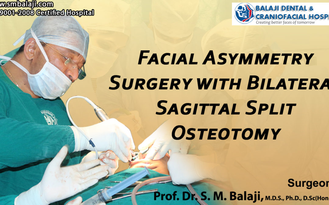
Under general anesthesia, incisions were first placed in the mandibular retromolar region bilaterally. Flaps were then elevated to expose the mandibular bone. Bone cuts were then made and bilateral sagittal split osteotomy was performed using the Obwegeser’s technique. The mandible was pushed posteriorly ensuring proper occlusion of the teeth.
This was then fixed in place using titanium plates and screws. Extreme care was taken throughout the procedure to ensure protection of the inferior alveolar nerve during mobilization of the proximal segment. All incisions were then closed with resorbable sutures.
Complete patient satisfaction with the outcome of the surgery
Results of the surgery were immediately evident to the patient and her parents. They were very happy with the outcome of the surgery. The patient’s occlusion was completely normal and she had an esthetic facial profile. She would need to undergo continued orthodontic treatment to bring her teeth into perfect alignment, thus obtaining good facial symmetry.
The patient expressed her happiness and said that this would transform her life completely.
The patient is a 21-year-old male from Kottayam in Kerala, India. He has had lower jaw asymmetry since his childhood. This had resulted in an asymmetrical face. There is however no history of any trauma or accidents to his jaw. This has been slowly becoming progressively worse throughout his growing years. There was also a component of open bite to his occlusion.
Chewing and speech problems had also increased exponentially with the passage of time. It has now reached a point where his lower face has become grossly deformed. This had affected him a lot and he had become socially withdrawn.
Problems in a social setting due to his facial deformity
This had led to considerable bullying in school and the patient had always been socially withdrawn. He had lost his self confidence due to his facial appearance and had very few friends. The patient’s parents stated that he was very depressed and they were worried about his well being.
They had taken him to a local oral and maxillofacial surgeon who had examined the patient. Realizing the complexity of the patient’s treatment needs, he had referred the patient to our hospital for management.
Our hospital is a specialty center for distraction osteogenesis surgery in India. Distraction osteogenesis is the technique used for bone lengthening. We were one of the first hospitals in India to utilize this method for jaw correction surgery. Patients with hemifacial microsomia are routinely treated for facial asymmetry.
Mandibular lengthening is routinely performed in our hospital through mandibular distraction osteogenesis. Our hospital is a referral center for many countries from South East Asia and Europe.
Initial presentation and treatment planning at our hospital
Dr SM Balaji, facial asymmetry surgeon, examined the patient and ordered for comprehensive imaging studies including a 3D CT scan. The right side of the patient’s mandible was longer than the left side. This had resulted in his gross facial asymmetry. The left side was noted to be deficient. There was also an obvious occlusal cant present.
Even though the defect was observed to be on the right side, it was decided that the patient needed to undergo treatment on the left side to correct the facial asymmetry. It was decided to utilize distraction osteogenesis on the left mandible to increase its length. A Univector mandibular ramus distractor was planned to be fitted on the left side.
This treatment was explained to the patient in detail who consented to surgery. The patient was scheduled to undergo facial asymmetry correction with distraction osteogenesis
Surgical correction of the facial asymmetry
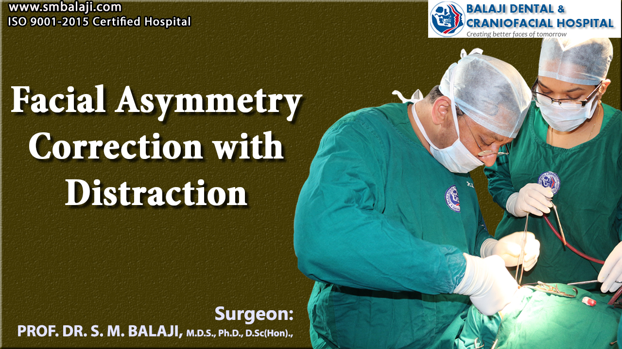
Under general anesthesia, an incision was made in the left mandibular retromolar region. A flap was elevated followed by bone cuts. A Univector mandibular ramus distractor was then fixed on the left side using titanium screws. Extreme care was taken throughout to ensure that the inferior alveolar nerve was not damaged during mobilization of the distal segment of the mandible.
Following successful fitting of the distractor, correction of the occlusal can was addressed next. A sulcular incision was placed in the maxilla followed by. Le Fort I bone cuts. The maxilla was then mobilized and the posterior end of the left maxilla was fixed using transosseous wires.
Hemostasis was achieved and closure was done using sutures. This was followed by Intermaxillary fixation using stainless steel wire. The patient was then taken to the recovery room in stable condition.
Postsurgical consolidation of bone before distraction
A latency period of about 6-7 days was allowed after surgery. Following the latency period, the distractor was activated with a distraction of 1 mm every day. A total of 13 mm of bone distraction was achieved over the next two weeks. There was symmetry of the two sides of the mandible at the end of this period.
A week later, a plate was fixed to the left posterior maxilla to prevent any further downward movement. The distractor was then removed after a period of about 3-4 months following bony consolidation at the site of mandibular distraction.
Successful outcome of the distraction osteogenesis procedure
The patient started noting the changes during the distraction phase itself. He was extremely happy at the end of the distraction phase of the treatment. This had resulted in good facial harmony through correction of his asymmetry. He stated that he was now ready to face life with a new found confidence.
No human face is truly symmetric. A mild degree of facial asymmetry is always present, but not easily spotted by the naked eye. Only when this asymmetry is easily noticeable does it require surgical correction. Distraction devices are commonly employed in the correction of facial asymmetry. The distraction devices are first fitted. This is followed by distraction. Consolidation of the distracted bone segment is allowed before removal of the distraction device. The bones heal and soft tissue remodeling occurs during this period of consolidation.
Correction of hemifacial microsomia and temporomandibular joint surgery also come under corrective jaw surgery. There are many surgical techniques that are employed for correction of facial asymmetry. All these are technique sensitive and are best performed by experienced surgeons.
Guidelines for surgical correction of orofacial deformities
The American Association of Oral and Maxillofacial Surgeons has laid down guidelines for all surgical procedures of the oral and facial regions. Bone grafts are advised in certain cases where distraction is contraindicated. Distraction osteogenesis and orthognathic surgery are two arms of facial asymmetry correction. Mandibular distraction osteogenesis is more common as mandibular asymmetry occurs more commonly than maxillary asymmetry.
Patient with a history of surgery for ameloblastoma
The patient is a 24-year-old male from Vandavasi in Tamil Nadu, India. He had developed a left lower jaw swelling around two years ago and visited a local hospital. Tests had been performed by an oral surgeon at the hospital. The patient had then been diagnosed with ameloblastoma of the left mandible. A left partial mandibulectomy had been performed followed by reconstruction with a rib graft and reconstruction plate. The patient had been left with a severe facial asymmetry with the right side of the mandible being longer than the left. This had depressed the patient to a degree that he refused to go outside. His career had suffered as a result and this alarmed his worried parents.
They had consulted with the oral surgeon again who realized the severity of the problem. The parents and the patient were informed that this could be addressed only at a specialist oral and maxillofacial surgery hospital. He had then referred them to our hospital for management of the patient’s facial asymmetry.
Our hospital is renowned for facial asymmetry surgery. Jaw reconstruction surgery is performed routinely at our hospital. Facial asymmetry resulting from trauma to congenital deformities of the jaws is addressed at our hospital.
Initial presentation at our hospital
Dr SM Balaji, jaw reconstruction surgeon, examined the patient and obtained a detailed history. The patient’s old medical records pertaining to the ameloblastoma surgery were studied in detail. He then ordered comprehensive imaging studies for the patient including a 3D CT scan. The patient was noted to have a deficient left mandibular body. Right side of the mandible was longer than the left. Mandibular ramus was also found to be defective.
Treatment planning presented to the patient and his parents
It was advised that the patient undergo mandibular ramus distraction osteogenesis on the left side. Removal of the reconstruction plate would be followed by mandibular ramus distractor fixation on the left side. The patient and his parents expressed understanding of the surgical procedure and consented to surgery.
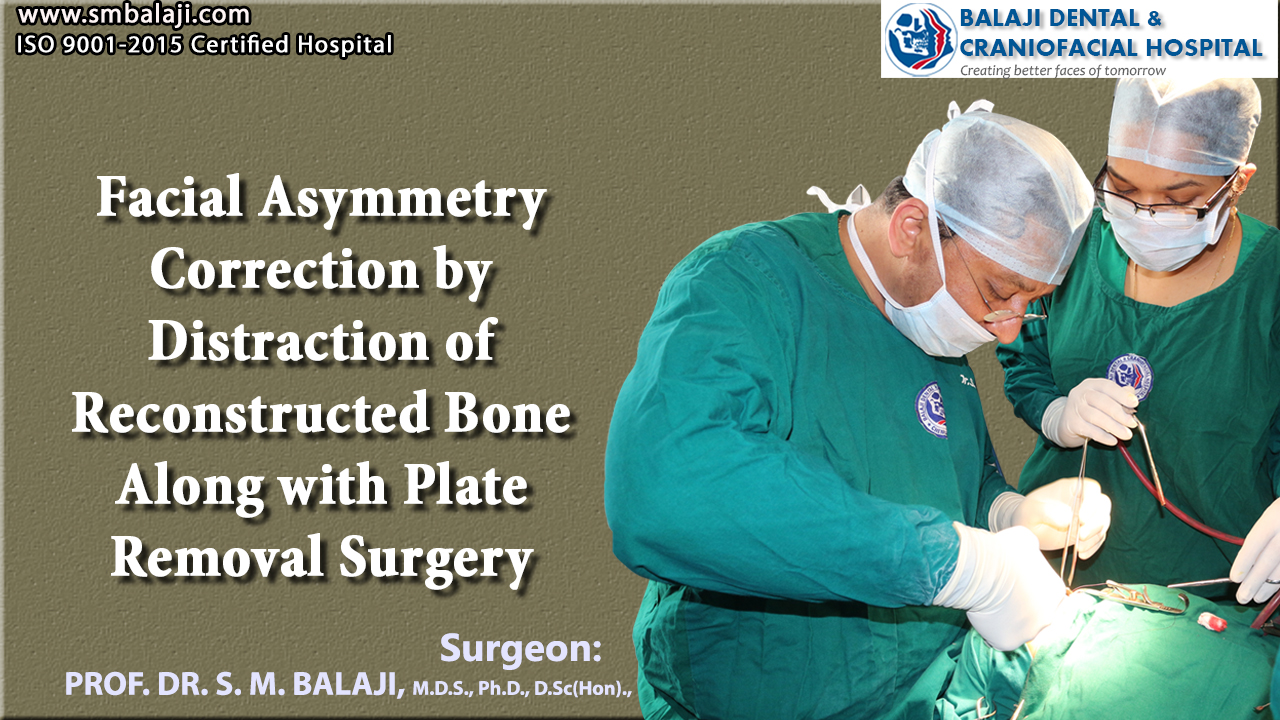
Successful surgical correction of facial asymmetry
Under general anesthesia, an incision was first made through the previous surgical scar in the left submandibular region. Dissection was done down to the mandibular reconstruction plate and screws. These were then removed. An incision was then made in the left mandibular retromolar region. A flap was elevated following which bone cuts were made. Mandibular ramus distractor was then fixed on the left side using titanium screws. The activating arm of the distractor was brought out through the left submandibular incision.
Following the fixation of the distractor in the mandible, a sulcular incision was made in the maxilla. Le Fort I bone cuts were then made and the maxilla was mobilized. The posterior end of the left maxilla was fixed using transosseous wires. Hemostasis was ensured following which closure was done using sutures.
Distraction osteogenesis of the mandible to establish facial symmetry
Interarch wiring was done following the surgery. A latency period of about 6-7 days was allowed for stabilization of the surgical site following which the distractor was activated. Two clockwise turns of the distractor were performed everyday for a resultant distraction of about 1 mm. A total of 18 mm distraction of the mandibular bone was achieved at the end of the treatment. A plate was then fixed to the left posterior maxilla to prevent further downward movement. The distractor will be removed after a period of about 3-4 months once adequate consolidation of the bone in the distracted segment is seen.
Complete patient satisfaction at the results
The patient started noticing the change during the distraction phase itself. Result was obvious at the completion of 18 mm of distraction. The patient was very happy with the outcome of surgery. He expressed his satisfaction at the final outcome of the surgery. The patient also added that he would be able to start leading a normal life again