Microtia is a congenital condition in which there in poor development of the ears. Correction involves staged reconstruction of the ear using autogenous costal cartilages. A template is utilized to create the form of the proposed external ear. Three surgeries complete reconstruction of the microtia affected ear.
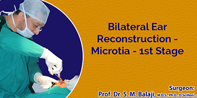
Patient presents to our hospital for specialized microtia surgery
This is a young 13-year-old boy from Tirupati, Telangana with microtia. His parents brought him to our hospital for bilateral microtia repair. Dr SM Balaji, microtia repair specialist, explained the surgery to them. They agreed to proceed with surgery.
Microtia surgery of bilateral ears performed for young boy from Tirupati
After general anesthesia, rib grafts were first harvested. Grafts obtained were from the fourth, fifth and sixth ribs. A Valsalva maneuver was then performed. This was to rule out accidental perforation into the thoracic cavity. Using a metal template, the sixth rib graft was first carved and sculpted to form the external ear framework. This was done for both the right and the left sides. The other graft pieces were then fixed and secured with nonresorbable sutures. Care was taken to maintain symmetry between the frameworks of both ears.
The skin of the right ear was first incised and underlying tissues dissected to create a pocket. The framework of ribs was then tunneled into the pocket. Skin was then sutured and surgical drain placed at the site. This step will help adapt the skin to the cartilage framework. The left ear was then addressed.
Second stage repair will be after about 3 months. This will be for elevation of the framework inserted in the first stage.
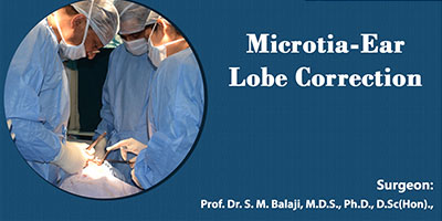
Stage 2 microtia correction after successful stage 1 microtia surgery
The patient is now 14 years old. He has already undergone stage 1 costal cartilage placement under the skin. This had recreated the form of the left pinna. Stage 2 surgery involves lifting up the reconstructed left pinna.
This lies flat against the side of the head after the stage 1 surgery. His left ear lobe was also deformed with skin tags. This too needed corrective surgery. He presents now for stage 2 surgery.
Lifting up of reconstructed external ear from side of head
Under general anesthesia, an incision was first made around the reconstructed pinna. This was then lifted up and stabilized in position with sutures. Attention was then turned to the deformed ear lobe. A skin incision was first made in the ear lobe and adapted to give normal form to the deformed lobe. Sutures were then used to close the incision.
The patient expressed his happiness at the results before discharge from the hospital.
Ear Reconstruction Surgery(Microtia) for a Seychelles boy
9-year-old Nathan Parkash from Seychelles was born with completely absent external portion of his right ear, he only had a very small, rudimentary ear lobule. His left ear was normal. Doctors in his hometown diagnosed that the child’s hearing was normal. As the boy started going to school, his social interactions increased, children at school began teasing him because of his ear. He suffered ridicule and unwelcome stares. He saw that he was not able to wear sunglasses and walkmans like his fellow mates. He started becoming psychologically depressed about his appearance. Slowly he became withdrawn and self-conscious. His parents were becoming increasingly worried for their child’s poor self-esteem. They looked for hospitals that do ear reconstructions all around the world and they found that such centers are very few and the results were not up-to the mark. Usually people from Seychelles go to South Africa or Dubai for medical treatment. They found that very few hospitals were available for doing such procedures but the outcomes were not satisfactory.
The Govt. of Seychelles referred them to our hospital for the ear reconstruction surgery (Microtia) as they found the surgical results of the surgeon to be exceptionally best in India. Dr. S.M. Balaji decided to perform complete ear reconstruction in 2 stages using the boy’s own rib graft (costochondral graft) as artificial materials can lead to inherent problems like infection, allergy and loss of stability over time. Using the patient’s own graft is the best technique giving permanent and stable results.
The first stage was called cartilage-banking surgery. Based on the measurements of the normal left ear, a template was made. It was important to do this correctly as the ear characteristics have to be accurately replicated to achieve symmetry. A costochondral graft was taken from the rib. Using the template as reference, the graft was contoured to create the structural framework. This is most crucial and has to be done with great precision and accuracy. The skin of the right ear was raised creating a pocket and the cartilage framework was placed below the skin. Additional piece of cartilage was also placed adjacent for future use. A surgical drain was placed that created a suction making the overlying skin to adapt to the framework and the ear shape was achieved
After 3 months the second stage surgery called ear-lifting surgery was done. Using the excess preserved cartilage and skin graft, the entire ear framework was lifted from the side of the head giving a normal, raised appearance to the ear. Following reconstruction the ear was given a more aesthetic, natural-looking appearance. As the boy grows the normal left ear will grow, but the reconstructed right ear will not increase in size. So to avoid re-surgery, the right ear was reconstructed with a higher dimension matching the adult size.
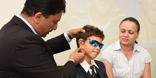
Following second stage surgery the boy is delighted about his “new ear”. Also he is happy that he won’t be teased any longer. He and his parents are happy with the surgery outcome and returned to their hometown. He may require revision surgery at later stage. Ear reconstruction surgeries are challenging and must be performed with skill, care & artistic sense. Timely correction of ear defects is important for the person’s physical and emotional well-being.