Mandibular prognathism Correction (Long Lower jaw) Surgery BSSO Bilateral Sagittal Split Osteotomy

Mandibular prognathism Correction (Long Lower jaw) Surgery BSSO Bilateral Sagittal Split Osteotomy

Mandibular prognathism arising from an excessively large mandible

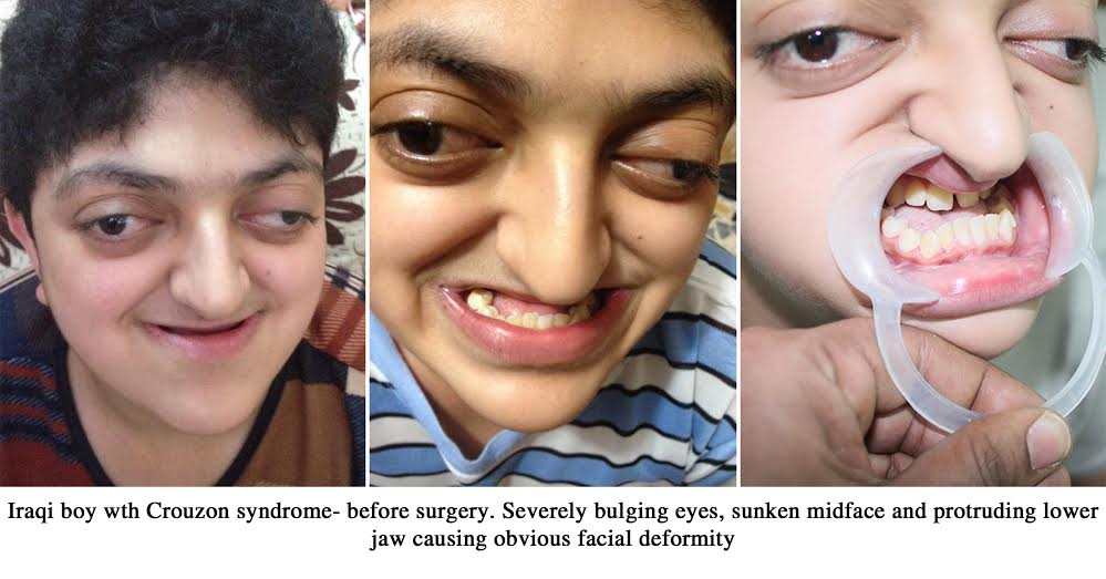
When the lower jaw is disproportionately larger than the upper jaw in size, this condition is known as mandibular prognathism. When the upper jaw is normal in size and the lower jaw is disproportionately larger than normal, it is called true mandibular prognathism. Correction of this condition involves reduction in the size of the mandible. This is achieved through surgical correction.

Benefits from the development of modern medicine and dentistry to humanity

Modern dentistry has come to the aid of many conditions that caused great suffering in the past. There was a time when even the most minor of dental ailments could even turn life threatening. Alveolar abscesses that are so easily controlled today through the use of antibiotics and root canal treatment could lead to the death of the individual 150 years ago.

Mandibular prognathism during olden days would have caused an extreme degree of distress to the individual with regards to eating and speech. Anterior crossbite is present in mandibular prognathism. Difficulty with chewing food would have caused intake of insufficient nutrition that could lead to malnutrition.

Historical occurrence of mandibular prognathism

Mandibular prognathism was a feature that was common among the von Habsburgs of Austria. The Habsburgs were the dynasty that ruled the Austro-Hungarian empire before World War I. Most of the male members of this dynasty demonstrated a marked mandibular prognathism. A prognathic mandible is also called a Habsburg jaw or lantern jaw.
The Habsburg jaw was an extreme manifestation of mandibular prognathism because of many generations of inbreeding. What must have been present as mild mandibular prognathism in the first generation had become accentuated to an extreme degree through the inbreeding. It was so extreme in some of them that they were unable to chew food because of the prognathism. There was no treatment to correct jaw prognathism prior to the advent of modern dentistry.
Jaw deformities are a common occurrence in case of extensive inbreeding. Many breeds of dogs that are inbred to retain the bloodline exhibit severe jaw deformities. This is because inbreeding is against the laws of nature and is something that is unique to human beings.

Surgical technique used for correction of mandibular prognathism

A bilateral sagittal split osteotomy is performed to reduce the size of the mandible and bring it into correct alignment with the maxilla. An illusion of mandibular prognathism can occur when there is a retruded maxilla and a normal mandible. Correction of this condition is through forward movement of the maxilla through distraction osteogenesis. This corrects the relationship between the maxilla and mandible and brings the two jaws into correct alignment.

Surgeons advice diagnosis or treatment planning in several steps using various diagnostic protocols. They look for excessive wear of the teeth in the molar region. This surgery is contraindicated in patients who have undergone treatment for oral cancer as per the American Association of Oral and Maxillofacial Surgeons.

Patient with mandibular prognathism referred to our hospital for surgical correction

The patient is a young woman from Kurnool, Andhra Pradesh. She has had long standing problems with anterior crossbite due to mandibular prognathism. This had led to her feeling very self conscious because of the cosmetic aspect of her prognathism. She had always desired to undergo corrective jaw surgery for her problem.
Parents and patient referred to our hospital for surgical management

Her parents decided to seek the upper and lower jaws and teeth correction treatment advice from an oral surgeon in their hometown. She and her parents approached a local oral surgeon to seek the advice regarding the details about surgery. He said to them that this was not conducive for maintaining good oral health and referred them to Balaji Dental and Craniofacial Hospital for corrective orthognathic surgery. He explained to them that this is a specialty maxillofacial surgery center for jaw reduction surgery in India. Jaw reduction surgery requires extreme precision in measurements to achieve the best results.

Mandibular prognathism is a condition that is present in 0.35% of the Indian population. This condition completely alters the jaw line. Correction of this condition is done by jaw reconstruction surgeon in India. A bilateral sagittal split osteotomy is performed to reduce the size of the lower jaw and make it proportionate with the upper jaw.

Benefits of undergoing surgical correction of mandibular prognathism

It is common to see many people with mandibular prognathism who have not even considered the option of surgical correction. They have to be educated regarding the benefits of undergoing surgical correction of their condition. This would include improvement in speech and eating habits along with the improvement in esthetics. It is mostly those with pronounced functional disruption who voluntarily seek help for their condition.

Tooth extraction is never a part of this surgery as it is performed posterior to the tooth bearing section of the mandible. This can also be considered a cosmetic surgery or plastic surgery as it results in dramatic improvement in facial appearance of the patient. Soft tissues automatically mold themselves once this surgery is complete and there is no need to do any soft tissue correction as part of this surgery.

Initial examination and treatment planning explained to the patient and her parents

Dr SM Balaji, oral and maxillofacial surgeon, examined the patient and ordered radiographic studies. The patient had anterior and right-sided posterior crossbite. Presurgical planning was meticulously carried out on the patient. He explained that orthognathic surgery is performed for correction of this condition. The patient was in agreement with the treatment plan and consented to surgery.

Surgical correction of mandibular prognathism with very good results

Under general anesthesia, bilateral sagittal split osteotomy was performed. The anterior segment of the mandible was then positioned posteriorly to align correctly with the maxilla. Great care was taken to protect the exposed inferior alveolar nerve during this segment of the surgery. Intermaxillary fixation was then applied. This was followed by removal of a 5 mm wide piece of bone from the horizontal plane bilaterally followed by fixation of the two segments with plates and screws. Occlusion was checked again and maxilla and mandible were found to be in perfect alignment. Intermaxillary fixation was then removed and the patient was extubated and recovered uneventfully from general anesthesia.

Surgery Video

Successful cleft lip repair for unilateral cleft lip

Successful cleft lip repair for unilateral cleft lip

Baby girl from Mathura   

This little baby girl from Mathura was born with a cleft. She is now 3 months of age. Mathura accounts for a higher than average number of babies born with a cleft. Her upper lip was split into two. This is known as a cleft lip. She also had a hole in the roof of her mouth which affected her feeding. The patient’s mother was also operated for a cleft lip and palate as an infant. An ultrasound test during pregnancy revealed the baby’s cleft. There was also a familial history of clefts running through the generations.

Unilateral cleft lip

Cleft lip is a congenital split in the upper lip on one side often associated with cleft palate. The cleft palate usually involves the soft palate and hard palate. Babies with cleft lip usually have difficulty in feeding. Cleft babies may develop various problems as they continue to grow. They have dental problems which require corrective treatments. The dental problems require surgical and non-surgical intervention. They have an increased risk of middle ear infections which may lead to hearing problems. Babies born with clefts may have speech problems. Thereby requiring a speech therapist opinion.

Cleft lip surgeon in India

 Though aware of the condition the parents were very depressed. They were very concerned about her future. They were searching far and wide for the best cleft lip surgeon in India. A local physician referred them to our hospital. Dr.S.M.Balaji one of the leading cleft lip and palate surgeon in India examined the patient. He planned to correct the cleft lip at 3 months of age.

Primary cleft lip repair

Corrective surgery to repair the lip is required. Cleft lip and palate surgeon Dr. S.M. Balaji planned to perform primary lip repair at 3 months of age. Unilateral cleft lip repair is done using Modified Millard’s technique. The upper lip musculature is also corrected during the surgery.

Surgery outcome

The result of the surgery was as expected. She looked like any other baby girl of her age with minimal to no scar. The parents were very pleased with the results.

Future surgical corrections

Cleft palate repair will be done at about 9 months of age. Bone grafting is to be done at 3 and a half to 4 months of age. Speech correction / Pharyngoplasty may be necessary at 3-4 years of age. Further surgical corrections will be carried out at later date. The mouth palate must be repaired within 10 months of age.

TMJ reconstruction surgery with costochondral graft for hemifacial microsomia

TMJ reconstruction surgery with costochondral graft for hemifacial microsomia

Hemifacial microsomia and the manifestations of the condition

Hemifacial microsomia is a congenital condition where one side of the face is underdeveloped with the eyes, ears, cheekbone and mandible being affected. The lower half of the face is affected the most by this condition. Underdevelopment of the mandible includes underdevelopment of the TM joint. Hemifacial microsomia always results in TMJ disorders. This can result in serious structural and functional disorders of the TMJ. One manifestation of this condition is extreme facial asymmetry. Normal alignment between the upper and lower jaws is also lost.

Other symptoms of hemifacial microsomia include an extremely wide mouth, skin flap present over an underdeveloped external ear and growths around the eye on the affected side.

Normal and abnormal relationship between the upper and lower jaws

Normal alignment of the teeth is called normal occlusion. Normal occlusion of the teeth signifies normal alignment of the maxilla and the mandible. Normal alignment of the jaws can also be present in cases of abnormal relationship between the teeth of the upper and lower jaws. This is called dental malocclusion and correction of this is through fixed orthodontic treatment. Dental malocclusion can also result when there is abnormal alignment between the two jaws. This is known as skeletal malocclusion. Skeletal malocclusion can only be corrected through surgery. Surgery that is performed to correct the relationship between the two jaws is known as orthognathic surgery.

Manifestations and etiology of hemifacial microsomia

TMJ symptoms begin to manifest early in life for these patients. There is also a lot of TMJ pain. Most cases of hemifacial microsomia occur as the result of a combination of genetic and environmental factors. It causes serious problems with the patient’s general health as well as oral health. Relaxation techniques do not help with pain arising from this condition as it is caused by a structural deformity. Clinical trials are constantly being performed around the world to help alleviate the symptoms in the long term.

The temporomandibular joint is the only movable joint in the skull. It is the point of contact between the mandible and the skull. The joint has a cartilaginous capsule that provides synovial articulation between the glenoid fossa of the temporal bone and the mandible. There are a variety of disorders that lead to disruption in the functioning of this joint.

Fractures of the TMJ are very common as any blow to the chin is directly transmitted to the joint. The condylar fractures of the mandible are amongst the most common facial fractures. Only nasal bone fractures occur in greater numbers. Common causes of condylar fractures are road traffic accidents and interpersonal assaults.

Classification of disorders of the TMJ

There are many conditions that lead to disorders of the TMJ. They can be broadly classified into congenital, traumatic, idiopathic, degenerative and inflammatory disorders. Congenital disorders include absence of the joint at birth, smaller than normal or larger than normal joint and abnormally developed joint.

Traumatic disorders include dislocation, subluxation and fracture. An idiopathic disorder of the TMJ is defined as one where there is pain and dysfunction of the joint without any identifiable cause for the symptoms. Inflammatory disorders include myositis, capsulitis and synovitis. Degenerative disorders include rheumatoid and osteoarthritis.

Benefits of surgery in patients with temporomandibular joint disorders

Oral and maxillofacial surgeons always recommend surgery for this condition. There is overall improvement in the patient’s health once surgery is performed. Physical therapy in the form of jaw exercises has to be performed regularly to maintain good joint health following surgery. Surgery for hemifacial microsomia however is not an orthognathic surgery or corrective jaw surgery. This can be categorized under temporomandibular disorders that require TMJ reconstruction surgery.

Patient with hemifacial microsomia with worsening mandibular deviation

The patient is an 8-year-old girl with right-sided hemifacial microsomia. She also has microtia of her right ear and underdevelopment of her mandible with deviation to the right side. She had first undergone right-sided jaw reconstruction surgery elsewhere when she was 3 years old. However, over the course of time, her right-sided mandibular deviation had gotten worse with development of an open bite and her parents consulted with a plastic surgeon.

Referral to our hospital by a plastic surgeon in her hometown

The plastic surgeon examined the patient and explained to the parents that the patient needed TMJ surgery for temporomandibular joint reconstruction, which came under Oral and Maxillofacial Surgery. He explained that the American Association of Oral and Maxillofacial Surgeons (AAOMS) had developed a protocol for surgical treatment of this condition. Only certain hospitals in India met the stringent standards prescribed by the AAOMS for TM joint surgery.

The patient was thus referred by him to Balaji Dental and Craniofacial Hospital. It was explained to the parents that the cost of temporomandibular joint reconstruction surgery in India was a fraction of what it cost in other countries with an excellent infrastructure for healthcare.

Examination and treatment planning at our hospital

Dr SM Balaji, an experienced temporomandibular joint reconstruction surgeon, examined the patient. He then ordered a 3D CT scan and other pertinent imaging studies. This protocol is standard for determining the best treatment option for the patient. These diagnostic studies revealed that the patient had extreme resorption of the costochondral rib grafts that had been placed during the previous surgery. Detailed treatment planning was done and it was decided to reconstruct the temporomandibular joint with rib grafts. This was explained to the parents of the patient in detail and they consented to the proposed treatment plan.

Dr. SM Balaji is an experienced TMJ surgeon who has published many articles on the jaw joint surgeries performed by him in many international scientific journals. Many cases of hemifacial microsomia have been surgically rehabilitated at our hospital. Postsurgical follow up of over ten years has shown excellent results with the patient leading active lives that were fully integrated into their society.

Harvesting rib grafts for jaw reconstruction surgery

The patient was taken to the operation theater where she was prepped and draped for surgery. Under general anesthesia, two rib grafts were first harvested from the patient. These were harvested with the periosteum to ensure that the grafts would not resorb after reconstruction of the temporomandibular joint. A Valsalva maneuver was then performed to ensure that there was no perforation into the thoracic cavity. The incision was then closed in layers with sutures.

Jaw joint reconstruction using rib grafts

Attention was next turned to the right side of the mandible. A buccal vestibular incision was first made to expose the region of the mandible to be reconstructed. It was seen that the rib grafts used to reconstruct the mandible in the previous surgery had nearly fully resorbed. The rib grafts that had been harvested from the patient were then carefully shaped to attain the right anatomical dimensions of the patient’s jaw. These were then joined together using titanium plates and screws.

This was then placed in the region of the jaw to be reconstructed and fixed with titanium plates and screws to the right side of the mandible. Postoperative radiographs revealed perfect reconstruction of the right-sided temporomandibular joint, ramus and body of the mandible. The flap was then closed with sutures. Dental implants can be placed once the grafts have fully integrated with the reconstructed jaw.

The patient and her parents were very satisfied with the results of the surgery and expressed their gratitude before final discharge from the hospital. Instructions were given to the parents to return to the hospital in 3-4 years for a radiographic checkup of the reconstructed jaw joint.

Surgery Video

One stage microtia ear reconstruction surgery

One stage microtia ear reconstruction surgery

Microtia deformity correction surgery

The congenital underdevelopment of the external ear is defined as microtia. A completely undeveloped external ear is known as anotia. Anotia is the most extreme manifestation of microtia. Microtia is one of the birth defects that can appear in isolation or in conjunction with a syndrome. Some common syndrome that present with microtia include hemifacial microsomia, Treacher-Collins syndrome, Franceschetti syndrome and Goldenhar syndrome.

Prosthetic fabrication of external ears for microtia patients

Unilateral microtia is more common although occurrence of bilateral microtia is also recorded. The right ear is more commonly affected than the left ear. Fabrication of prosthetic ears using latex was in vogue in the early 1900-30s though esthetic results were not as satisfactory. Silicones are used currently for fabrication of prosthetic ears now and these ears offer satisfactory esthetic results for the patient. 

Studies to evaluate prospects of hearing ability

It is important to determine as early as possible if the patient’s hearing can be restored through surgery. A CT scan is performed at around the age of 5-6 to determine the degree of development of the external auditory canal. Placement of cochlear implants can be considered in cases of complete lack of development of the external auditory canal. This will restore normal hearing for the child.

Cosmetic aspects of microtia deformity

Microtia is cosmetically disfiguring for the patient and can lead to a lot of distress for the patient and his family. Such deformity correction is performed by ear deformity correction specialists. Correction of this deformity is through the use of rib cartilage grafts and is done in three stages when the deformity is severe. Preauricular skin grafting is utilized in cases of deficiency of skin cover for the rib graft

Modes of sound wave propagation

Sound waves are transmitted to the inner ear through two modes. One is air conduction and the other is bone conduction. What we know as normal hearing is through air conduction. This is much clearer and the sounds are much sharper. The sound waves are propagated through the external auditory canal and strike the ear drum in this case.

 The second mode of propagation of sound waves is through bony conduction. When we have a severe respiratory illness or an ear infection, the external auditory canal gets blocked. Though we are still able to hear sounds, it sounds muffled and has a vibratory quality. This is because the sounds we hear are reaching the eardrum through the bone. This is known as bony conduction.

Grades of microtia deformity

There are four grades of microtia. Grade I microtia features a less than complete development of the external ear. This external ear has a shape and form of a near normal external ear. A fully functional external auditory canal is also present. Grade II microtia features a partially developed ear where the top of the external ear is affected. This is informally known as a lop ear. It has a closed external auditory canal but the ability to hear is retained in these patients and it is through bony conduction of sound waves. Grade III microtia features a tiny remnant of the external ear that resembles an ear tag. This is the most common form of microtia. Grade IV microtia features the complete absence of an external ear.

Severe microtia is also accompanied by complete lack of an external auditory canal. This causes severe hearing impairment. It is very rare that a patient without an external auditory canal has the ability to ear through bony conduction.

It is very important that good blood supply is maintained when surgical reconstruction of microtia defect is performed. Gradual loss of blood supply to the cartilage graft over a period of time was the main reason for the failure of other microtia surgery technique. Loss of blood supply can lead to gradual withering of the reconstructed external ear.

Alternative techniques of microtia reconstruction

A few other reconstructive techniques were utilized for microtia reconstruction in the past although they have been discontinued to lack of good long term results. Treating microtia is also a form of plastic surgery. Cosmetic ear surgery requires the surgeon to have an artistic vision of the final surgical outcome. When the reconstruction is unilateral, the surgeon should be able to visualize the process through which symmetry of the ears will be achieved.

An ear framework template or cartilage framework is first constructed and the cartilaginous rib grafts that are harvested from the patient are crafted to suitable size and shape. The reconstructed ear should be symmetrical to the normal ear. This surgery can also be done in a single stage when the deformity is not extreme as illustrated in the case described below.

Young boy with microtia desires to undergo surgery

This is a young boy with microtia in the form of a congenitally deformed right external ear. He is now 8 years of age. Teasing at school by other children was causing great distress to him. His concerned parents consulted a local surgeon to enquire about ear reconstruction surgery in India. He explained to them that a good ear reconstruction surgeon would be able to correct his ear deformity.

Cost of microtia surgery in India

Cost of ear reconstruction surgery in India is only a fraction of what it costs in developed countries. The results are however as good as in any western country. He made extensive enquiries and referred them to Balaji Dental and Craniofacial Hospital. The patient and her parents subsequently presented at our hospital.

Patient presents at our hospital for microtia surgery

Dr SM Balaji, ear deformity correction surgeon, examined the patient. He ordered facial biometrics for both the deformed ear and the normal left ear.This would ensure that the reconstructed right ear was symmetrical to the normal ear. Biometric studies revealed this to be a constricted ear. Detailed planning for the surgery was done. Usual microtia ear reconstruction surgery is normally performed in three stages.He decided to do a single stage deformity correction as the microtia was not very severe. It was explained to the patient’s parents that a costocartilaginous rib graft needed to be harvested for the procedure. The patient’s parents were in agreement and consented to the surgery.

Surgical correction of microtia deformity

 Under general anesthesia, a costocartilaginous rib graft was first harvested from the patient. A Valsalva maneuver was next performed to ascertain that there was no perforation into the thoracic cavity. The incision was then closed in layers with sutures.

Attention was then turned to the deformed external ear. The harvested rib graft was then shaped to fit into the outer border of the helix of the external ear. Two incisions were then made in the helix and the cartilage tunneled through the incision to form the border of the ear helix. This resulted in a normal form to the external ear. A preauricular flap was raised to aid in full tissue coverage of the anterior incision. Both incisions were then closed with sutures.

The patient and her parents expressed complete satisfaction at the results of the surgery before final discharge from the hospital.

Surgery Video


Revision Rhinoplasty Surgery with Scar Removal in the Chest

Revision Rhinoplasty Surgery with Scar Removal in the Chest

Increasing numbers of revision rhinoplasty surgery in India

A revision rhinoplasty is performed when a patient is not satisfied with the results obtained from the original rhinoplasty surgery. It is also performed when there is a functional compromise from the original rhinoplasty. This could be worsening difficulty with breathing or snoring from a deviated nasal septum. A revision rhinoplasty is more difficult that a rhinoplasty as it has to offset the damage arising from the previous rhinoplasty. An inexperienced rhinoplasty surgeon might remove more bone or cartilage than necessary thus causing serious damage to the nasal structures.

The initial surgery is known as primary rhinoplasty and the revision rhinoplasty is known as secondary or corrective rhinoplasty.

Indications for rhinoplasty surgery or nose surgery

A rhinoplasty procedure or nose job is advised when a patient has breathing difficulties or excessive snoring during sleep. This can be due to a deviated nasal septum. A nasal septum deviation could be congenital or could be caused by trauma to the nasal bone. This results in a deviation in the nasal passage, thus obstructing the clear passage of air during breathing. A rhinoplasty procedure needs to be performed to rectify this. Rhinoplasty surgery restores the function of the nose back to optimal levels.

Principal qualities of a good rhinoplasty surgeon

An experienced rhinoplasty surgeon instinctively diagnoses the cause for the problem and the degree of correction required in each case. There is no unnecessary excision of tissue or removal of bone. This ensures that optimal results are obtained with maximum cosmetic as well as functional benefit to the patient from the surgery. It always results in an improvement in facial features.

Rhinoplasty surgery is not just purely a cosmetic surgery. It also results in relieving a person’s breathing difficulties. Facial plastic surgeons as well as oral and maxillofacial surgeons perform rhinoplasty procedures.

Different categories of nasal deformity corrected by rhinoplasty surgery

Rhinoplasty surgery is a form of facial plastic surgery. There are many kinds of nasal deformities that are addressed by a rhinoplasty. These include parrot beak deformity, flat nose deformity, hooked nose deformity, dorsal nasal hump, saddle nose deformity, broad nose deformity, crooked nose deformity and asymmetrical nose deformity. A rib cartilage is often used as a graft to enhance the bridge of the nose in case of a flattened nose. The protocol followed for rhinoplasty is the same as for other surgery procedures.

Cleft rhinoplasty is a highly specialized field. Only the most experienced of surgeons excel in this corrective procedure. It is a highly complex procedure and complete mastery over all the basic sciences of medicine is a prerequisite to master this.

Rhinoplasty surgery used to be confined to the rich and the famous in the past, but the number of rhinoplasty procedures has drastically increased leading to the increase in the number of hospitals offering this service to patients.

A rhinoplasty surgery requires extreme finesse and precision to give the best esthetic results. It takes years of training under an experienced surgeon to master this. When an inexperienced surgeon performs this surgery, chances of patient dissatisfaction with the results is high and this automatically leads to an increase in the number of revision surgeries being performed.

Patient dissatisfied with previous rhinoplasty surgery

The patient is a young girl who had undergone rhinoplasty elsewhere with placement of a rib graft to augment the bridge of her nose. She had also had placement of a graft to the tip of the nose. Over a period of time, she felt that the bridge of her nose was too broad and the tip of the nose was very pronounced. She desired to have a narrower bridge of the nose and a less pointy tip of the nose. She had presented to a plastic surgeon in her hometown to enquire about surgeons performing rhinoplasty surgery in India and also scar revision surgery in India. He referred her to our hospital. She therefore presented to our hospital requesting corrective surgery as it was a failed rhinoplasty and revision was advised.

Patient presents at our hospital for revision rhinoplasty

Dr SM Balaji, a well known rhinoplasty surgeon in India, examined the patient and ordered imaging studies. As the outcome of the initial rhinoplasty was not satisfactory to the patient, she was a little apprehensive and enquired about the revision rhinoplasty swelling timeline. It was explained to the patient that the recovery period would be for a period of one to two weeks and that she would need to stay home for the first ten days following surgery. The splint that would be placed on her nose after the procedure would be removed at this point.

He explained that the graft to the bridge of the nose would need to be shaped to a finer form during the revision rhinoplasty surgery. The patient also complained of an unsightly scar at the site of the bone graft harvest from the previous surgery. It was decided to perform a scar revision surgery at that site during this surgery. The scar revision procedure would involve closure of the wound in several layers for minimal residual scar formation.

Harvesting of bone graft for nasal bridge rhinoplasty augmentation

Under general anesthesia, a rhinoplasty incision was placed in the columella and the bone graft was taken out. It was then reshaped to form a finer bridge of the nose. It was placed again at the bridge of the nose. The tip of the nose was also shaped to be less pronounced. Incision was then closed with sutures.

Scar revision surgery performed with optimal results

Attention was then turned to scar removal from the chest wall. An incision was made at the old chest incision scar site. The unsightly scar tissue was excised completely. A Valsalva maneuver was then performed to ensure that there was no accidental perforation into the thoracic cavity. The incision was then closed in layers. The patient’s revision nose surgery was meticulously documented in her medical records.

Surgery Video


Facial Reconstruction Surgery of Upper Jaw after Treatment of Fungal Infection Followed By Dental Implant Surgery

Facial Reconstruction Surgery of Upper Jaw after Treatment of Fungal Infection Followed By Dental Implant Surgery

History of maxillary rhinosporidiosis with pain and drainage

This patient had been having undiagnosed pain and swelling for two years when he first came to our hospital. He had been referred here by a surgeon in his hometown after several doctors in various dental clinics had been unable to diagnose his condition. He even visited a few oral surgery implants clinics without any solution.

He was examined by Dr SM Balaji, oral and maxillofacial surgeon, who ordered a 3D CT scan and biopsy. Cultures of the biopsy specimen were done and revealed rhinosporidiosis infection of the maxillary sinus. Surgery was performed and all infected bone had been removed. This had lead to a maxillary bone deficiency.

A jaw reconstruction surgery was indicated for the patient. Jaw reconstruction surgery in India is very affordable compared to western countries. A jaw reconstruction surgeon in India undergoes years of rigorous training. The patient had lost most of his natural teeth in the maxilla and was partially edentulous. The jaw joint was however not compromised by this.

There were no retained tooth roots and the health of the soft tissues were not compromised. Only two wisdom teeth were present in his mouth, his left mandibular third molar, which was impacted, and his right maxillary third molar. Dental work following surgery would be the placement of implants followed by dental rehabilitation with crowns. The patient complained of excessive daytime sleepiness, but there was no suspicion of sleep apnea.

The patient was advised to come back to the hospital after adequate healing of the maxillary surgical site. Bony rehabilitation of the lower and upper jaws is also treated by plastic surgeons. Principles of orthognathic surgery are not used in these cases although this is also corrective jaw surgery.

Surgery for maxillary augmentation with bone grafts

The patient presented after the surgical wounds had healed completely. A biopsy obtained from the maxilla revealed complete resolution of his maxillary rhinosporidiosis. Augmentation of his maxillary bone had been performed with bone grafting for placement of dental implants at a later date. The patient presents now for placement of dental implants.

Nobel Biocare implants are very commonly used for dental implant surgery in India. Almost every implant surgeon in India uses this. Implant surgery is a component of maxillofacial surgery.

History of modern dental implants

It was Dr Per-Ingvar Branemark who first discovered osseointegration of titanium with bone. During the course of his research into factors that influence bone healing, he inserted titanium rods into rabbit leg bones. When he later tried to retrieve it, he discovered that the titanium rod had completely fused with the bone and the two had become indistinguishable. The structure of the bone was not affected in any way and bony strength to withstand stresses was also normal.

This later led to the development of dental implants by Dr. Branemark. He is considered to be the father of dental implantology. This led to the formation of Nobel Biocare and he continued to research ways to improve dental implants.

Advantages of dental implants over other dental prostheses

Titanium is very light and completely fuses with the bone. The joint between the bone and the titanium implant is virtually indistinguishable and very strong. It can withstand occlusal loads comparable to that of natural teeth. Constant research into dental implants has led to different implant designs to cater to individual patient needs.

The most common type of implant is the single tooth dental implant. The crown fixed to this type of implant can either be a single crown or a crown that is a part of a bridge. The number of implants used to fix a bridge depends upon various factors. Factors such as the amount of occlusal load the implant will have to bear determine this.

Different types of dental implants systems in use today

All on 4 implants utilize the placement of tilted dental implants for the rehabilitation of an entire arch. Placement of the teeth prosthesis, typically a bridge, can be done within 24 hours. It is very convenient for the patient and the postoperative recovery phase is the shortest for this implant. Patients thus rehabilitated are able to eat all varieties of food and the occlusal loads borne by these implants are equal to that of natural teeth.

Long-standing edentulous condition of the maxilla can lead to severe resorption of the maxillary bone. This can be to a degree that cannot support most implant systems. This lead to the development of zygoma implants. Dental implants are directly fixed to the zygomatic bone. The zygomatic bone is a very strong bone and serves as an ideal foundation to soak up occlusal forces that are exerted upon these implants.

Factors behind the long term success of dental implant surgery

The success rate of dental implants is very high. It ranges from 98% to 99.5%; however, it needs to be reiterated that the patient needs to take good care of the implants to enable long-term success of the implants. The habit of smoking greatly reduces the success of implant placement. The patient also needs to maintain meticulous oral hygiene with the use of aids such as dental floss and special mouthwashes that will be prescribed to the patient.

Maintenance of poor oral hygiene will definitely lead to failure of the implant system. The dental implant surgeon will have to ascertain the patient’s levels of motivation before going ahead with the placement of dental implants. Periodic checkup of dental implants needs to be done by the surgeon to ensure that the right conditions are being maintained in the oral cavity to ensure long term success of the dental implant treatment.

Placement of maxillary dental implants

A mucogingivoperiosteal flap was raised in the maxilla to expose the augmented maxillary bone. Screws used to fix the bone grafts to the maxilla during the previous surgery were removed. The bone grafts were seen to be fully integrated with the maxillary bone. Implants were then fixed in the maxilla. A total of six implants were used. Bio-Oss was then used to fill any remaining areas of a bony defect in the maxilla. The flaps were then closed with sutures.

Teeth will be attached to the implants once full osseointegration of the implants to the bone is complete. A dental implant surgeon in India usually waits for a period of six months before fixing crowns to the implants. The type of dental implants to be chosen is critical for the success of this surgery. The patient tolerated the procedure well and recovered uneventfully from general anesthesia.

Surgery Video