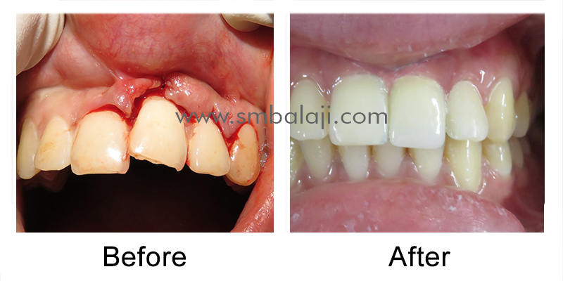
Patient with a history of unilateral cleft lip and palate repair
The patient is a 22-year-old from Dehradun in Himachal Pradesh, India who was born with a left sided unilateral cleft lip and palate. Lower facial growth is at times affected on the side of the cleft. Speech development is also affected if there is velopharyngeal incompetence. This is characterized by the presence of a hole in the roof of the mouth that creates a direct communication between the oral and nasal cavity. Speech therapy is needed after completion of the surgical phase of rehabilitation for complete normalization of speech.
Surgical repair of his cleft lip defect had been performed when he was 4 months old. This was performed at a local hospital. He had later undergone cleft palate repair when he was 10 months old. Cleft lip repair is ideally performed at 4 months of age and cleft palate repair is ideally performed at 10 months of age. He had later undergone alveolar cleft reconstruction at the age of 3-1/2 years. His teeth however erupted in a malaligned fashion due to his cleft defects. He had undergone fixed orthodontic correction of his malaligned teeth at the age of 12 years. His left lateral incisor was however congenitally missing due to the location of the cleft in the region of the left lateral incisor.
It had been advised to his parents that placement of a dental implant at that site would complete the rehabilitation process for him. He had undergone bone grafting with iliac crest to facilitate placement of the implant but there had been failure of the graft. A second bone graft surgery was attempted, but that too resulted in failure of the graft. He had been very disappointed by this and had settled for a removable denture to replace his missing left lateral incisor.
Referral to our hospital for bone graft surgery and dental implant placement
The patient had sought consultation at a few dental hospital regarding bone graft surgery, but they had expressed their inability to perform that surgery. It was then that an oral surgeon in Nainital had informed the patient that this procedure could be performed successfully at a specialty cleft lip and palate repair hospital and had referred the patient to our hospital for treatment
Initial examination and treatment planning at our hospital
Upon arrival at our hospital, Dr SM Balaji, cleft lip surgery and cleft palate surgery specialist, examined the patient and obtained a detailed oral history. A 3D CT scan was obtained to study the region of the bony cleft in detail. This revealed deficient alveolar bone in the region of the left lateral incisor. The patient also had malocclusion of his teeth despite undergoing fixed orthodontic treatment elsewhere.
It was explained to the patient and his parents that he first needed to undergo fixed orthodontic treatment first so that sufficient space could be created for placement of the implant. This would be followed by bone graft placement at the site of the bony deficiency to enable placement of the dental implant. This bone graft would be harvested from the mandibular symphyseal region as the previous grafts from the iliac crest and rib region had been rejected. The patient and his parents were in agreement with this treatment plan and the patient began fixed orthodontic treatment. Sufficient space was created after six months of treatment for placement of the implant and the patient is now ready for bone graft placement.
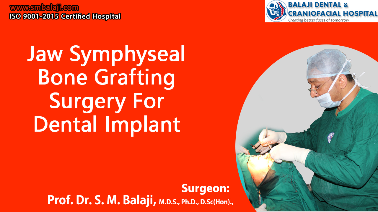
Successful placement of bone graft in the maxillary bone
Under general anesthesia, a crevicular incision was made in the anterior mandible and a flap was elevated to expose the symphyseal bone. Bone cuts were made in the region and a strip of buccal cortical bone was harvested from the region. Following this, a midcrestal incision was placed in the maxilla in the region of the left upper lateral incisor and a flap was elevated. The area of the bone defect was then exposed and reconstructed using the bone graft, which was fixed in place using titanium screws. Closure of the incision was done using resorbable sutures.
Implant surgery planning
The graft was perfectly placed in the region of the bony defect in the maxilla. A period of three to four months would be allowed for the graft to successfully blend in with the maxillary bone following which dental implant surgery would be performed. A further period of six months would be allowed for complete osseointegration of the implant with the bone following which a crown will be placed on the implant to complete rehabilitation of the patient.
A crown is an artificial tooth that is fabricated to mimic a natural tooth and is fixed on top of the implant. These artificial teeth are fabricated from ceramics or zirconium. Implant surgery can also be performed under local anesthesia in cases without any complications.
The patient and his parents were extremely happy with the results of the surgery and were instructed to report back to the hospital in three to four months for dental implant surgery.
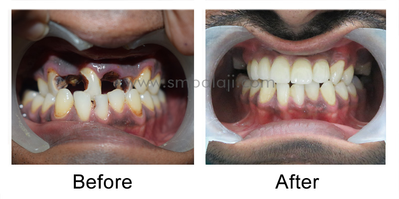
[vc_section content_layout=”full” animation_delay=”” disable=”” id=”” class=”” bg_type=”image” bg_image=”” color_overlay=”” enable_parallax=”” enable_pattern=””][vc_row content_layout=”boxed” equal_height=”” animation_delay=”” disable=”” id=”” class=”” bg_type=”image” bg_image=”” color_overlay=”” enable_parallax=”” enable_pattern=””][vc_column layout=”normal” vertical_align=”top” animation_delay=””][vu_heading style=”2″ heading=”CASE REPORT” subheading=”” alignment=”left” custom_colors=”” class=””][vc_column_text]This is a case of a 32 year old male patient. He approached our Dental Hospital hoping to find a solution for his long standing dental defect. The patient complained that his upper front teeth were broken, discolored and unpleasant aesthetically. He expressed that his neglect towards oral hygiene over the period of years, affected his teeth and compromised his oral functions to a great extent.
Also, the patient grumbled upon his difficulty to chew food, as his upper posterior teeth were also in a bad state of condition. He expressed his desperate need to fix his teeth, as it affected his self confidence and well being.
[/vc_column_text][vu_heading style=”2″ heading=”EXAMINATION” subheading=”” alignment=”left” custom_colors=”” class=””][vc_column_text]On clinical examination, the patient’s upper anterior and posterior teeth were grossly decayed, that only the root portion of the teeth structure remained, which cannot be conserved. The lower jaw teeth were in the initial stages of decay, due to the poor oral care of the patient. A full mouth x-ray (OPG) taken shows, badly cavitated and damaged teeth in the upper jaw.[/vc_column_text][vu_heading style=”2″ heading=”TREATMENT PLAN” subheading=”” alignment=”left” custom_colors=”” class=””][vc_column_text]After complete examination of the patient, considering the patients need and age, Dr. SM Balaji, Maxillofacial surgeon and Implantologist, decided to extract the badly decayed teeth and root stumps, followed by Dental Implant placement under local anesthesia. Patient was completely satisfied with the treatment plan. Consent was obtained[/vc_column_text][vu_heading style=”2″ heading=”DENTAL IMPLANT PLACEMENT ” subheading=”” alignment=”left” custom_colors=”” class=””][vc_column_text]Dr.S.M.Balaji extracted the defective teeth in the upper jaw that cannot be preserved by means of any dental treatment, under local anesthesia. Subsequently, the gum tissue surrounding the relative site were raised exposing the underlying jaw bone. Dental implants of appropriate height were fixed in the bone. A total number of five teeth were removed in the upper anterior region, yet, a set of three implants has been fixed at the relative site, which will later be covered by the loading of a fixed ceramic bridge onto the implants.
This was done to make the treatment cost effective for the patient. Closure of the elevated gum tissue is achieved with absorbable suture. Patient was asked to report after a duration of 3 months. This healing period is required for the dental implants to completely integrate with the jaw bone. Until then, a provisional prosthesis was given to the patient, to replace his missing teeth.[/vc_column_text][vu_heading style=”2″ heading=”THE MAKEOVER” subheading=”” alignment=”left” custom_colors=”” class=””][vc_column_text]The patient came back after 3 months. Post operative OPG taken shows well positioned dental implants, fully consolidated with the jaw bone. Final measurements were taken and bite trials seen. Later, ceramic bridge of exact color match to his natural teeth were fixed to the implants. The patient was very happy to have a natural-looking smile and to be able to bite and speak well.[/vc_column_text][/vc_column][/vc_row][/vc_section][vc_row content_layout=”boxed” equal_height=”” animation_delay=”” disable=”” id=”” class=”” bg_type=”image” bg_image=”” color_overlay=”” enable_parallax=”” enable_pattern=””][vc_column layout=”normal” vertical_align=”top” animation_delay=”” width=”1/2″][vc_single_image image=”5708″ img_size=”full” add_caption=”yes”][/vc_column][vc_column layout=”normal” vertical_align=”top” animation_delay=”” width=”1/2″][vc_single_image image=”5709″ img_size=”full” add_caption=”yes”][/vc_column][/vc_row][vc_row content_layout=”boxed” equal_height=”” animation_delay=”” disable=”” id=”” class=”” bg_type=”image” bg_image=”” color_overlay=”” enable_parallax=”” enable_pattern=””][vc_column layout=”normal” vertical_align=”top” animation_delay=”” width=”1/2″][vc_single_image image=”5710″ img_size=”full” add_caption=”yes”][/vc_column][vc_column layout=”normal” vertical_align=”top” animation_delay=”” width=”1/2″][vc_single_image image=”5711″ img_size=”full” add_caption=”yes”][/vc_column][/vc_row]
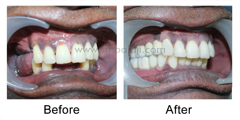
[vc_section content_layout=”full” animation_delay=”” disable=”” id=”” class=”” bg_type=”image” bg_image=”” color_overlay=”” enable_parallax=”” enable_pattern=””][vc_row content_layout=”boxed” equal_height=”” animation_delay=”” disable=”” id=”” class=”” bg_type=”image” bg_image=”” color_overlay=”” enable_parallax=”” enable_pattern=””][vc_column layout=”normal” vertical_align=”top” animation_delay=””][vu_heading style=”2″ heading=”PATIENT FROM VELLORE” subheading=”” alignment=”left” custom_colors=”” class=””][vc_column_text]A 56 year old male patient from Vellore, Tamil Nadu, India, approached our Dental Hospital seeking for a permanent replacement of his missing teeth. The patient had allegedly, got his upper and lower posterior teeth removed, due to extensive decay, at his native a few years back. The patient had been experiencing great difficulty to bite and chew food properly. Patient had been using a removable denture for the past 1year to hide his dental defect. He lacked self confidence and was hesitant to smile.[/vc_column_text][vu_heading style=”2″ heading=”COMPLAINT” subheading=”” alignment=”left” custom_colors=”” class=””][vc_column_text]The patient complained that, the denture often got displaced on talking and chewing. On the other hand, there has been recurrent ulcers in his mouth due to long hours of wearing the denture. Also, he was not able to taste and relish his meals as the upper denture covered the roof of his mouth. This had altered the patient’s standard of living and appearance. Patient demanded for a fixed option that would last for a lifetime and, significantly, improve his well-being.[/vc_column_text][vu_heading style=”2″ heading=”ANALYSIS OF THE PATIENT” subheading=”” alignment=”left” custom_colors=”” class=””][vc_column_text]Clinically, there were missing posterior teeth in the upper and lower jaw, with missing anterior teeth in relation to the lowers. Preoperative OPG taken revealed lack of posterior teeth in the upper and lower jaw. On the other hand, there was sufficient bone height with regard to the relative site, which makes him an ideal candidate for a fixed treatment option.[/vc_column_text][vu_heading style=”2″ heading=”TREATMENT PLAN” subheading=”” alignment=”left” custom_colors=”” class=””][vc_column_text]In view of the patient’s need and demand, maxillofacial surgeon and Implantologist, Dr. SM Balaji planned to replace the missing teeth with Dental Implants under local anesthesia, followed by a fixed prosthesis downstream. Patient’s medical history affirmed no abnormalities. Consent obtained from patient to move on with the treatment plan.[/vc_column_text][vu_heading style=”2″ heading=”TREATMENT PROCEDURE” subheading=”” alignment=”left” custom_colors=”” class=””][vc_column_text]Under local anesthesia, the gum tissue surrounding the corresponding site is cut and elevated, exposing the underlying jaw bone. Dental Implants of appropriate size are placed in the bone with stability and retention. Once the implants are in place, the surgical site is thoroughly flushed with saline. The gum tissues were then approximated with dissolvable suture. A course of antibiotics and painkillers were prescribed to the patient.[/vc_column_text][vu_heading style=”2″ heading=”FOLLOW-UP” subheading=”” alignment=”left” custom_colors=”” class=””][vc_column_text]A temporary prosthesis had been given for the patient’s use for the time being. A duration of 3 months was required for the dental implants to completely osseointegrate with the teeth bearing bone. Until then, the patient continued on with the provisional prosthesis. Post operative x-ray taken showed well positioned, integrated, Dental implants.
Post-satisfactory healing, final impression was taken and bite registration was done. A natural looking ceramic prosthesis was fixed onto the dental implants. The prosthesis instantly gave him a makeover. The patient was so happy that, he couldn’t stop smiling![/vc_column_text][vc_row_inner equal_height=”” animation_delay=”” disable=”” id=”” class=””][vc_column_inner vertical_align=”top” animation_delay=”” width=”1/3″][vc_single_image image=”5685″ img_size=”full” add_caption=”yes”][/vc_column_inner][vc_column_inner vertical_align=”top” animation_delay=”” width=”1/3″][vc_single_image image=”5686″ img_size=”full” add_caption=”yes”][/vc_column_inner][vc_column_inner vertical_align=”top” animation_delay=”” width=”1/3″][vc_single_image image=”5687″ img_size=”full” add_caption=”yes”][/vc_column_inner][/vc_row_inner][vc_row_inner equal_height=”” animation_delay=”” disable=”” id=”” class=””][vc_column_inner vertical_align=”top” animation_delay=”” width=”1/3″][vc_single_image image=”5688″ img_size=”full” add_caption=”yes”][/vc_column_inner][vc_column_inner vertical_align=”top” animation_delay=”” width=”1/3″][vc_single_image image=”5689″ img_size=”full” add_caption=”yes”][/vc_column_inner][vc_column_inner vertical_align=”top” animation_delay=”” width=”1/3″][vc_single_image image=”5690″ img_size=”full” add_caption=”yes”][/vc_column_inner][/vc_row_inner][vc_row_inner equal_height=”” animation_delay=”” disable=”” id=”” class=””][vc_column_inner vertical_align=”top” animation_delay=”” width=”1/3″][vc_single_image image=”5691″ img_size=”full” add_caption=”yes”][/vc_column_inner][vc_column_inner vertical_align=”top” animation_delay=”” width=”1/3″][vc_single_image image=”5692″ img_size=”full” add_caption=”yes”][/vc_column_inner][vc_column_inner vertical_align=”top” animation_delay=”” width=”1/3″][/vc_column_inner][/vc_row_inner][/vc_column][/vc_row][/vc_section]
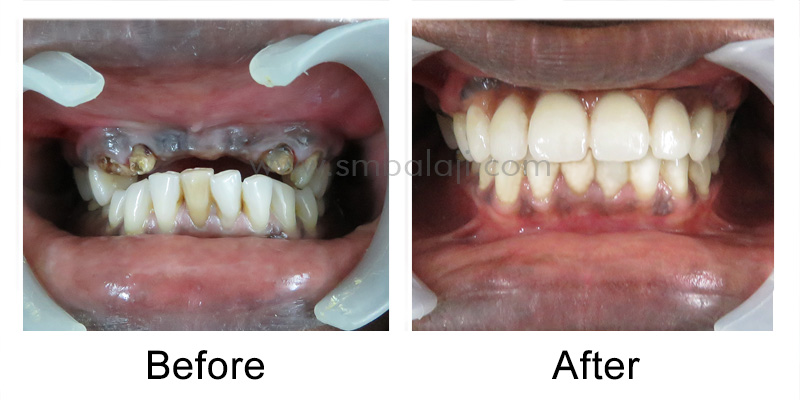
[vc_section content_layout=”full” animation_delay=”” disable=”” id=”” class=”” bg_type=”image” bg_image=”” color_overlay=”” enable_parallax=”” enable_pattern=””][vc_row content_layout=”boxed” equal_height=”” animation_delay=”” disable=”” id=”” class=”” bg_type=”image” bg_image=”” color_overlay=”” enable_parallax=”” enable_pattern=””][vc_column layout=”normal” vertical_align=”top” animation_delay=””][vu_heading style=”2″ heading=”PATIENT FROM ANDHRA PRADESH” subheading=”” alignment=”left” custom_colors=”” class=””][vc_column_text]A 32 year old school teacher from Andhra Pradesh, India approached our Dental Hospital seeking for a fixed replacement of her missing teeth. The patient gives a history of road traffic accident 5 years back. She experienced a traumatic fall which had knocked out her upper front teeth. In order to hide her dental defect, for the past 3 years patient was using the dental bridge given elsewhere after root canal treatment of the adjacent teeth.[/vc_column_text][vu_heading style=”2″ heading=”COMPLAINT” subheading=”” alignment=”left” custom_colors=”” class=””][vc_column_text]The patient complained that the dental bridge often got dislodged which hampered her speech and function. This eventually led to bad breath due to food getting stuck in between the bridge and the gums. Also, the patient stated that there was intermittent pain in her upper front teeth which increases on chewing. This affected the patient’s quality of life. She felt too timid to speak and smile. She was in need of a permanent solution to get back her good old smile.[/vc_column_text][vu_heading style=”2″ heading=”EXAMINATION ” subheading=”” alignment=”left” custom_colors=”” class=””][vc_column_text]On clinical examination, the dental bridge exhibited mobility. There was impaction of food and debris in between the bridge and the gums. The mobile dental bridge was removed and the condition of the teeth were evaluated. The tooth structure-supporting the dental bridge were broken and weak. Pre-operative full mouth x-ray taken indicated missing upper central incisors. There was incomplete root canal treated teeth on either side supporting the dental bridge owing to the pain stated by the patient earlier.[/vc_column_text][vu_heading style=”2″ heading=”TREATMENT PLAN” subheading=”” alignment=”left” custom_colors=”” class=””][vc_column_text]Treatment options like re-RCT of the teeth followed by traditional bridge was explained to the patient. However the patient was concerned and was not willing to go through the same process again. Also she was doubtful of its longevity. Hence considering the patient’s age and demand for a permanent fix, Dental Implants were opted. Dr.S.M Balaji, renowned Maxillofacial Surgeon and Implantologist, intended to place dental implants after extraction of the failed root canal treated teeth under local anesthesia. Patient’s consent was obtained.[/vc_column_text][vu_heading style=”2″ heading=”DENTAL IMPLANT PLACEMENT” subheading=”” alignment=”left” custom_colors=”” class=””][vc_column_text]Under local anesthesia, Dr SM Balaji extracted the broken teeth on either side of the upper arch. Following this, the gum tissue surrounding the corresponding site was raised exposing the underlying bone. Dental Implants of appropriate height were then placed in the empty tooth socket with stability. The implanted site was thoroughly irrigated with saline solution and closed with dissolvable suture.
The patient was given medications for 5 days to cope with the mild swelling which eventually subsided. During the intervening time, a self cleansing provisional prosthesis was given to replace the missing teeth.[/vc_column_text][vu_heading style=”2″ heading=”POST TREATMENT FOLLOW-UP ” subheading=”” alignment=”left” custom_colors=”” class=””][vc_column_text]It took a course of 3 to 4 months for the Dental Implants to completely integrate and harmonize with the jaw bone. Post operative OPG taken revealed well positioned dental implants in chime with the jaw bone. Subsequently, final bite trials were done. A natural looking, well-fitting fixed prosthesis was placed onto the implants in the upper jaw. The patient’s look elevated instantly. Her insecurities ceased! The prosthesis blended well with the adjacent natural teeth. She walked out with a dazzling smile! Patient is on a regular follow-up.[/vc_column_text][vc_row_inner equal_height=”” animation_delay=”” disable=”” id=”” class=””][vc_column_inner vertical_align=”top” animation_delay=”” width=”1/3″][vc_single_image image=”5664″ img_size=”full” add_caption=”yes”][/vc_column_inner][vc_column_inner vertical_align=”top” animation_delay=”” width=”1/3″][vc_single_image image=”5665″ img_size=”full” add_caption=”yes”][/vc_column_inner][vc_column_inner vertical_align=”top” animation_delay=”” width=”1/3″][vc_single_image image=”5666″ img_size=”full” add_caption=”yes”][/vc_column_inner][/vc_row_inner][vc_row_inner equal_height=”” animation_delay=”” disable=”” id=”” class=””][vc_column_inner vertical_align=”top” animation_delay=”” width=”1/3″][vc_single_image image=”5667″ img_size=”full” add_caption=”yes”][/vc_column_inner][vc_column_inner vertical_align=”top” animation_delay=”” width=”1/3″][vc_single_image image=”5668″ img_size=”full” add_caption=”yes”][/vc_column_inner][vc_column_inner vertical_align=”top” animation_delay=”” width=”1/3″][vc_single_image image=”5669″ img_size=”full” add_caption=”yes”][/vc_column_inner][/vc_row_inner][/vc_column][/vc_row][/vc_section]
[vc_section content_layout=”full” animation_delay=”” disable=”” id=”” class=”” bg_type=”image” bg_image=”” color_overlay=”” enable_parallax=”” enable_pattern=””][vc_row content_layout=”boxed” equal_height=”” animation_delay=”” disable=”” id=”” class=”” bg_type=”image” bg_image=”” color_overlay=”” enable_parallax=”” enable_pattern=””][vc_column layout=”normal” vertical_align=”top” animation_delay=””][vu_heading style=”2″ heading=”COMPLAINT ” subheading=”” alignment=”left” custom_colors=”” class=””][vc_column_text]A 21 year old young medical student from Chennai rushed to our Dental Hospital with lacerated gum tissue and uprooted upper left front tooth. Patient reported to have had an accidental fall at her house a couple of hours back. The traumatized area was profusely bleeding, also the patient was in immense pain. The pain was so awful that the patient requested on an immediate solution to bring her out of the agony.[/vc_column_text][vu_heading style=”2″ heading=”CLINICAL & RADIOLOGICAL EXAMINATION ” subheading=”” alignment=”left” custom_colors=”” class=””][vc_column_text]On clinical examination, there was a significant amount of swelling on the patient’s upper lip. The upper left front tooth had been displaced and mobilized. Also the gum tissue surrounding the relative site had been teared from the traumatic fall.
OPG (full mouth x-ray) taken revealed horizontal root fracture of the upper left front tooth. No pathological findings seen on the surrounding tooth structure. No signs of bone fracture.[/vc_column_text][vu_heading style=”2″ heading=”TREATMENT PLAN” subheading=”” alignment=”left” custom_colors=”” class=””][vc_column_text]Treatment option of stabilizing the fractured tooth with a rigid wire followed by observation of the tooth for 3 to 4 weeks was explained to the patient. As the patient was young and conscious esthetically, she demanded immediate correction of the fractured teeth. She requested for a permanent solution as she was planning to attend an upcoming event in a couple of months.
Considering the patient’s age and need, renounced maxillofacial surgeon and Implantologist, Dr. SM Balaji decided to extract the fractured tooth followed by immediate dental implant placement under local anesthesia. The patients age and adequate bone level was ideal for a dental implant as the bone tends to repair rapidly when compared to a much older patient. A thorough blood investigation revealed no underlying abnormalities. The treatment plan was explained and patient’s consent was obtained.[/vc_column_text][vu_heading style=”2″ heading=”IMMEDIATE DENTAL IMPLANT PLACEMENT” subheading=”” alignment=”left” custom_colors=”” class=””][vc_column_text]Under local anesthesia, Dr. SM Balaji extracted the fractured upper left front tooth. The lacerated gum tissues were raised. Dental implant of adequate height was carefully placed with precision. The gum tissues were cleaned with saline and closure was achieved with dissolvable suture. Post operative OPG shows well positioned implant at the relative site.[/vc_column_text][vu_heading style=”2″ heading=”TREATMENT FOLLOW UP” subheading=”” alignment=”left” custom_colors=”” class=””][vc_column_text]The patient was under antibiotics and painkiller for 3 days to cope up with the moderate swelling and discomfort. Patient had to wait for a healing period of 3 months for the bone to osseointegrate with the implant. Meanwhile a removable prosthesis was given to the patient as it would look aesthetically unpleasant without upper front teeth.
Eventually after a duration of 3 months, a fixed ceramic prosthesis was placed over the implant. The ceramic prosthesis blended well with the adjacent teeth giving her a natural alluring look. The patient was extremely happy with the outcome . She walked out with a ravishing smile. Patient had been on regular follow up over a period of 3 years[/vc_column_text][vc_row_inner equal_height=”” animation_delay=”” disable=”” id=”” class=””][vc_column_inner vertical_align=”top” animation_delay=”” width=”1/2″][vc_single_image image=”5630″ img_size=”full” add_caption=”yes”][/vc_column_inner][vc_column_inner vertical_align=”top” animation_delay=”” width=”1/2″][vc_single_image image=”5631″ img_size=”full” add_caption=”yes”][/vc_column_inner][/vc_row_inner][vc_row_inner equal_height=”” animation_delay=”” disable=”” id=”” class=””][vc_column_inner vertical_align=”top” animation_delay=”” width=”1/2″][vc_single_image image=”5632″ img_size=”full” add_caption=”yes”][/vc_column_inner][vc_column_inner vertical_align=”top” animation_delay=”” width=”1/2″][vc_single_image image=”5633″ img_size=”full” add_caption=”yes”][/vc_column_inner][/vc_row_inner][/vc_column][/vc_row][/vc_section]