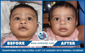
Patient born with left-sided cleft lip, palate and alveolus deformity
The patient is a 19-year-old male from Thanjavur in Tamil Nadu, India. The product of a consanguineous marriage, he was born with a unilateral cleft lip, palate and alveolus deformity. His parents were referred to a dental surgeon in the hospital who explained the treatment protocol to them. They had then been referred to our hospital for management.
Upon arrival at our hospital, an examination was performed followed by a comprehensive treatment plan. He had subsequently undergone cleft lip surgery at 3 months and cleft palate surgery at 8 months. His cleft alveolus deformity had been addressed with a bone graft at 7 years. He had been under the care of an orthodontist from the age of 10 onwards.
Development of maxillary retrusion with a resultant sunken face appearance
As the patient grew up, he noticed that his maxilla was getting retruded. This was caused by a hypoplastic maxilla, which resulted in a sunken face deformity to his face. There was also the gradual development of a skeletal anterior crossbite. This had resulted in speech and eating difficulties.
There was also a degree of compromise to his facial esthetics. He felt that it made him look abnormal. This was causing him to be socially withdrawn and depressed.
The patient discussed this with his parents and they decided to return to our hospital to address this. Our hospital is a premier center for facial cosmetic surgery in India. Our hospital has always been at the forefront of advancements in orthognathic surgery in India.
Scores of patients have benefitted from the facial esthetic surgery performed in our hospital. Reconstructive facial plastic surgery is also performed in our hospital for the facial region. Sunken cheeks are usually filled out when orthognathic surgery is performed for retruded jaws.
Patient and his parents present for correction of his sunken maxillary defect
Dr SM Balaji, Le Fort I surgery specialist, examined the patient and obtained imaging studies including a 3D CT scan. The patient had a skeletal anterior crossbite. It was explained to the patient that he needed to undergo maxillary advancement surgery to correct this problem. The patient and his parents expressed understanding of the treatment plan and consented to undergo surgery.
Successful surgical correction of his midfacial maxillary deformity
Under general anesthesia, a vestibular incision was made in the maxillary sulcus. A Le Fort I osteotomy was then performed and the maxillary bone disjointed. The maxillary bone was then advanced forward and occlusion was checked.
This was followed by fixation of the maxillary bone with titanium plates and screws. The incision was then closed with sutures. Anesthesia was successfully reversed and the patient was taken to the recovery room in stable condition.
Complete patient satisfaction with the results of the surgery
There was an immediate improvement in the patient’s facial esthetics following surgery. He now had a pleasing facial profile following forward advancement of his maxilla. The patient and his parents said that he would now develop more self confidence. They expressed their thankfulness before final discharge from the hospital.