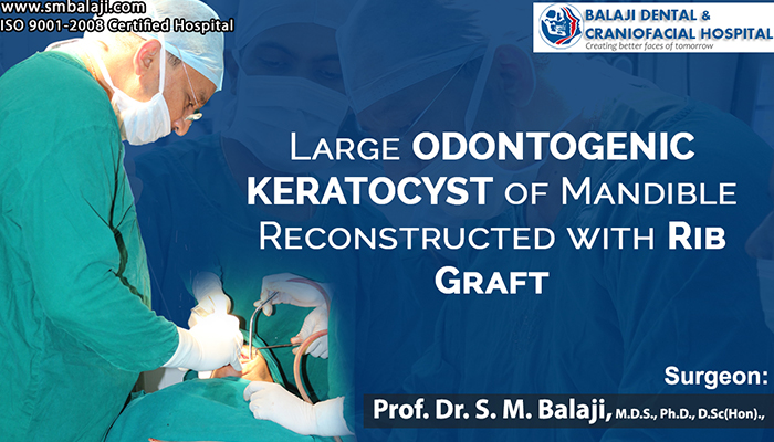
Genesis and characteristics of an odontogenic keratocyst
A cyst is essentially a sac of membranous tissue that can occur anywhere in the body. They normally contain fluid, but other substances can also be found inside them on occasion. Cysts are benign and not cancerous growth. There are many kinds of cysts. They include epidermoid cyst, sebaceous cyst, pilonidal cyst, ovarian cyst, chalazion of the eyes, popliteal cyst and pilar cyst amongst others.
Varieties of common cystic lesions
Treatment of cysts includes excision and careful enucleation of the cystic lesion including the membranous lining of the cyst. Any remnants left behind during enucleation can lead to recurrence of the cyst. Care has to thus be taken to ensure complete removal of the contents of the cystic cavity. Some of the more common cysts include the sebaceous cyst, the chalazion, and the epidermoid cyst. Cysts can turn painful when they occur in a confined space or get infected.
Epidermoid cysts are slow growing cysts that are the result of keratin buildup under the skin. They can get infected easily as they are very close to the surface of the skin. Sebaceous cysts occur when sebum glands get clogged leading to a buildup of sebum. This too can get infected easily. Surgical excision is the treatment of choice for both these cysts.
A pilonidal cyst occurs at a hair follicle and is said to occur due to a combination of hormonal changes, friction or prolonged pressure to that region. It can be quite painful and there is a foul smelling discharge from the cyst. A hair follicle is also present in association with the cyst. Treatment is curettage and enucleation along with removal of the associated hair follicle.
Etiology and pathogenesis of odontogenic keratocyst
An odontogenic keratocyst is a very rare benign developmental cyst that is very aggressive. It results in extensive destruction of the bone. It is most commonly seen in the posterior mandibular region in the third decade of life. The PTCH1 gene, which leads to the occurrence of odontogenic keratocyst has also been linked to the occurrence of ovarian cysts and ovarian cancer. Differential diagnoses for odontogenic keratocysts can include epidermoid cysts though these are completely different in their origin.
Recommended treatment protocol for odontogenic keratocysts
Treatment of the odontogenic keratocyst involves meticulous resection to completely remove the lesion followed by reconstruction of the jaw with bone grafting. Implant surgery for the placement of dental implants is performed after full bony consolidation of the bone grafts to complete full oral rehabilitation for the patient. This is the treatment protocol that is recommended by the American Association of Oral and Maxillofacial Surgeons. The patients thus properly cared for can go on to lead a completely normal life.
Use of dental implants for oral rehabilitation from destructive jaw lesions
The advent of dental implant treatment has enabled complete rehabilitation patients with odontogenic keratocyst. Implants enable replacing missing teeth. Success rates are extremely high for patients rehabilitated with dental implants. This is because dental implants mimic tooth roots and are able to bear occlusal loads that are borne by natural teeth.
Before dental implant treatment became a part of routine surgical protocol, postsurgical dental rehabilitation was through the use of removable dentures. This was highly unsatisfactory for the patient. The patient faced a lot of difficulty with both chewing and speech.
Dental implants have enabled the complete rehabilitation of both the upper and lower jaws. Proper maintenance of dental implants aided by following instructions of the implant surgeon meticulously is essential for the success of dental implant treatment.
Patient develops pain and swelling in the left posterior mandibular region
The patient is an 18-year-old female who had slowly developed a soft tissue swelling of the left posterior mandible with pain for the last six months. She had consulted a local dentist who noticed that the patient’s left third molar was missing from the oral cavity. Suspecting the swelling to be a dentigerous cyst, he had referred the patient to our hospital for management. Our hospital is a renowned center for jaw reconstruction surgery. Implants will need to be placed to complete oral rehabilitation after jaw reconstruction surgery.
Examination of the patient at our hospital with subsequent investigations
The patient presented at our hospital for management of the pain and swelling in her left posterior mandibular region. Dr SM Balaji, an oral and maxillofacial surgeon and jaw reconstruction surgeon in Chennai, examined the patient and ordered imaging studies and a biopsy of the lesion. The biopsy results returned as odontogenic keratocyst.
Imaging studies revealed a radiolucent lesion in relation to the left mandibular molars and a horizontally impacted third molar. Treatment planning for the management of the odontogenic keratocyst was explained to the patient in detail. She was advised to undergo cyst removal surgery and was in total agreement with surgical management of the lesion.
Bone graft harvested from the patient for jaw reconstruction
Under general anesthesia, rib grafts were first harvested from the patient. The rib grafts will be used to reconstruct the jaw after resection of the odontogenic keratocyst. A Valsalva maneuver was then performed to ensure that there was no perforation into the thoracic cavity. Following this, the incision was then closed in layers with sutures.
Resection of the odontogenic keratocyst from the left posterior mandible.
A mucogingivoperiosteal flap was raised in the left posterior mandibular region. This exposed the area of the odontogenic keratocyst. The cystic lesion was exposed and then completely resected. Great care was taken to ensure that there were no cystic remnants left behind in the bony cavity. The rib grafts were then carefully shaped to fit into the bony defect left behind by the lesion.
Titanium screws were used to fix the rib grafts into the bony defect in the jaw to reconstruct the jaw. Once adequate jaw reconstruction had been achieved with the rib grafts, the flap was then closed with sutures. The healing process along with bone remodeling of the grafts to merge in with the mandibular bone takes up to 6-8 months. Implants should only be placed after this is complete to ensure good long term results.
Placement of dental implants for complete oral rehabilitation for the patient
Once adequate consolidation of the bone grafts has been achieved, the patient will return to our hospital again for placement of dental implants. A period of six months will be given following dental implant surgery to allow for complete osseointegration of the dental implants with the mandibular bone. Complete oral rehabilitation of the patient would then be done with placement of tooth coloured crowns to the dental implants. Zirconium crowns or ceramic crowns offer great functional and esthetic results to complete the patient’s rehabilitation.
This young man is from Chennai, Tamil Nadu. He had a bike accident a week ago. Direct impact to his mandible resulted in a fracture of the mandible. He sustained facial injury as a result of this accident. This resulted in inability to close his mouth with an open bite. There were no soft tissues injuries from this accident. The patient never lost consciousness. He remained lucid during the immediate period after the fracture. Examination by a neurologist revealed no signs of head injury in the patient.
The neurologist explained to the family that the helmet had saved the patient’s life. He explained through charts how the injuries would have been very severe if the patient had not been wearing his helmet.
Presentation at our hospital for management of fracture
His family wanted the best treatment for his jaw fracture. They made enquiries about the best jaw fracture surgeon in India. He was then brought to our Balaji Dental and Craniofacial Hospital for treatment. Our hospital is a premier hospital for jaw fracture surgery in India. Success rate of surgery for mandibular fractures at our center is amongst the best in India. Our hospital is a specialty maxillofacial surgery center. We deal with cases of maxillofacial trauma on a daily basis. Our center is a top referral center among city plastic surgeons.
Special training through workshops are a regular feature at our hospital. Many oral and maxillofacial surgeons undergo this training. Fractures of the bones of the face are a common feature at these workshops. Injuries to the face are a common occurrence in the city. Treating these injuries needs the utmost care.
Treatment plan presented and consent obtained from patient
Dr SM Balaji, facial trauma surgeon in India, examined the patient. He obtained imaging studies for the patient. There was no fracture involvement of the eye sockets. There was no involvement of other facial bones or soft tissue. Any dental implants along the fracture line would need removal if present. Fracture was only at the left mandible. This came under the classification of facial fractures. There had been no facial lacerations from the accident. Location of the fracture determined his treatment plan.
Rigid fixation was essential for fracture stability. Fracture treatment would be through open reduction and internal fixation. This decision was based upon his experience with jaw fractures correction. The patient consented to the treatment plan. All appropriate consents were next signed by the patient and surgery scheduled.
Open reduction versus closed reduction
Open reduction and closed reduction are two ways of setting a fractured bone. The fractured segments of the bone stay reduced when it is a favorable fracture. The anatomy of the fracture ensures this. Certain fractures can be reduced without any skin incisions. These stay in place without displacement with plaster casts alone. This is a closed reduction. The break has to be clean without comminution of the fracture.
Fractures that do not stay reduced need open reduction and internal fixation. An incision is first made to gain access to the fracture site. Titanium plates and screws are then used to fix the fragments of bone to each other. This results in stabilization of the fracture. Incisions used to access the fracture are then closed with sutures. This is then followed by a period of immobilization for consolidation of bone. A closed reduction is possible only in a favorable fracture. All other fractures need open reduction and internal fixation.
Fractures of the mandible can be favorable or unfavorable fractures. Favorable mandibular fractures stay stabilized with closed reduction and intermaxillary wiring. Care should be taken to maintain proper occlusion of the teeth. These fractures heal without any further intervention.
Unfavorable mandibular fractures can comprise of several fracture segments. These do not stay stabilized with closed reduction. They need correction through stabilization with titanium plates and screws. An incision is first made to access the fracture site. The fracture fragments are then brought together into proper anatomical alignment. Titanium plates and screws are then used to stabilize the fracture. Occlusal harmony of the teeth should be ensured before final closure. The patient needs to return for periodic checks for a prescribed period of time. Full bone consolidation at the fracture site ensures complete healing of the fracture. The number of plates used for fracture reduction would increase with fracture severity.
Successful open reduction and internal fixation of the fracture
Under general anesthesia, a left vestibular incision in the mandible exposed the fracture. The fracture segments were then brought into correct alignment and occlusion checked. The fracture was then stabilized with plates and screws. Incisions were then repaired by suturing.
The patient expressed his satisfaction at the results of the surgery before discharge. He was able bring his teeth together. The open bite had undergone complete resolution. Facial esthetics was also perfect and there was no residual asymmetry.
Patient with deficient maxilla presents for augmentation surgery
The patient is a middle aged man from Waltair. He had undergone an endoscopic surgery for clearance of maxillary sinus rhinosporidiosis. A complete maxillary resection was performed previously at our hospital to remove all affected bone and bone affected by osteomyelitis. A reconstruction was done using the remaining bone. This resection however led to a maxillary bone deficiency, causing problems with nutrition and speech. He was then sent for a course of medical treatment of his rhinosporidiosis with complete resolution of his infection. He then presented to our hospital for definitive management of his problems.
Rhinosporidiosis treated with full resolution
Dr SM Balaji, facial reconstruction specialist, examined the patient. A biopsy was first obtained from the mucosa. Once it was confirmed that there was complete resolution of his fungal infection, the patient was then scheduled for surgery.
Maxillary augmentation surgery performed with bone grafts
Under general anesthesia, a rib graft was first harvested from the patient. A Valsalva maneuver was then performed to confirm patency of the thoracic cavity. The incision was then closed in layers.
Successful completion of maxillary augmentation surgery
Attention was next turned to the maxilla. A mucoperiosteal flap was then raised and plates from the previous surgery removed. Pieces of rib graft were then fixed at the deficient regions. This aided in augmenting the deficient maxillary bone. Once adequate augmentation was performed, the flap was then closed using sutures. Implants at a later date will complete oral rehabilitation of the patient.
The patient expressed his happiness at the progress of his treatment. He expressed his gratitude at the successful completion of the first phase of treatment.
This is a 32-year-old man from Palakkad. He complained of severe pain and swelling in his lower jaw. He also complained of difficulty opening his mouth and chewing food. He has been suffering from this condition for about four years now. He searched the Internet for the best jaw correction surgeon. His search led him straight to our hospital.
Dr SM Balaji examined the patient. He recommended comprehensive testing for the patient. Clinical, radiological and histopathological examinations were then performed. This led to the diagnosis of an odontogenic keratocyst. There was complete obliteration of the mandibular body.
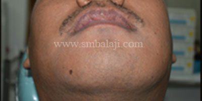
Preoperative facial profile showing huge swelling in the lower half of the face
Preoperative facial view showing huge swelling in the lower half of the face
OPG showing extensive bone loss involving the body of the mandible
3DCT showing extensive loss of buccal cortex
Costochondral rib graft harvested to reconstruct the affected portion of mandible
Costochondral graft site immediately after suturing
Infected portion of mandible removed
Cystic lesion along with the affected bone and teeth removed in toto
Harvested rib graft used to reconstruct the entire body of the mandible using Ti plates and screws
Suturing after surgical reconstruction of lower jaw
Treatment plan included complete cyst resection and reconstruction of the lower jaw. This would be by utilizing costochondral grafts. The affected area of the lower jaw was then removed. This was along with the involved tooth to ensure zero recurrence. The costochondral rib grafts were then used to reconstruct the lower jaw. Titanium screws and plates helped achieve stable fixation of the grafts . The patient was very happy with the outcome of the surgery. There was no scar as all incisions were intraoral.
Dr. S.M. Balaji explains the advantage of implants to the patient:
This patient presented at Balaji Dental and Craniofacial Hospital, Teynampet, Chennai, a few months ago with multiple missing teeth in both jaws. The patient has been partially edentulous for many years now. The patient requested dental implants to replace the missing teeth. Dr SM Balaji examined the patient and ordered CBCT(Cone Beam CT Scan) diagnostic studies in order to do bone mapping of the patient’s jaws. It was found that bone had resorbed in both the maxilla as well as the mandible.
It was explained to the patient that the existing bone was inadequate for fixing the implants and that bone grafting would be required to reconstruct the maxillary and mandibular alveolar ridges in order to place the implants. The patient consented to surgery and bone grafting was performed.
Implants placed for the patient:
The patient presents now for placement of dental implants in the reconstructed areas of the maxilla and the mandible. Midline alveolar ridge incisions were made and the reconstructed areas of the bone were exposed. Screws that were fixed to hold the grafts in place were removed since the grafts had fused to the alveolar bone. Implants were placed in the reconstructed regions of the maxilla and the mandible.
The alveolar flaps were then closed with sutures. Once adequate osseointegration had taken place between the implants and the bone over a period of 4-6 months, the flaps would be raised again and the crowns would be fixed on the implants.