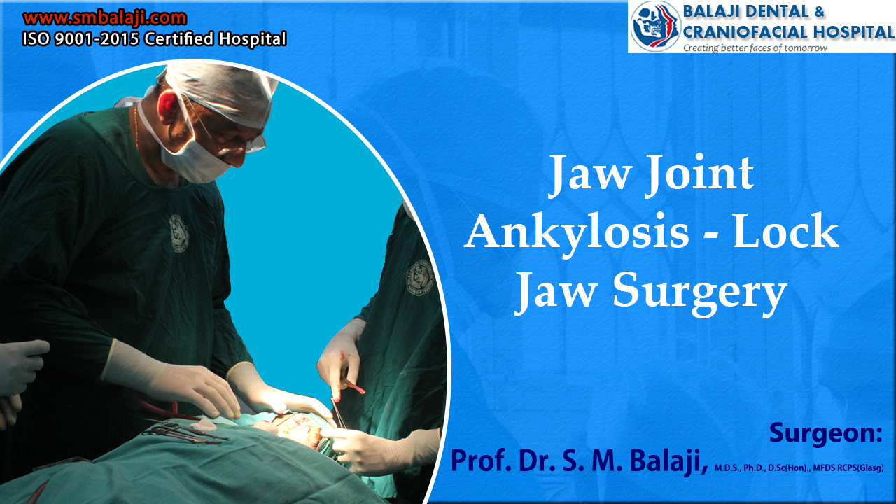
Patient with inability to open her mouth for many years now
The patient is a 10-year-old girl from Rajahmundry in Andhra Pradesh, India. She had extremely limited mouth opening of about 2-3 mm for many years now. This has made it nearly impossible for her to eat any solid food. The patient had been diagnosed with temporomandibular joint ankylosis many years ago.
She has been on a liquid diet for a long time and had lost a lot of weight. The patient rarely plays with other children as her speech was also affected. She also exhibited very poor social skills because of her problem. Her parents were greatly worried about her overall health. There was also an anterior open bite.
Deciding to get this addressed, they had taken her to a nearby city for consultation. An x-ray at a hospital had revealed that she had ankylosis of her right temporomandibular joint. They had counseled the parents extensively and had referred them to our hospital for management of the patient’s condition.
Our hospital is a renowned center for temporomandibular joint ankylosis surgery. Hundreds of patients have been successfully rehabilitated here after lock jaw surgery. Temporomandibular joint reconstruction surgery is also routinely performed in our hospital.
Initial presentation at our hospital
Dr SM Balaji, jaw reconstruction surgeon, examined the patient and ordered pertinent imaging studies including a 3D CT scan. This revealed that there was complete bony ankylosis of the right temporomandibular joint. The patient also had extremely restricted mouth opening and a retruded mandible.
Her parents were not able to recall any trauma that could have led to the ankylosis. It was advised that she has to undergo surgical correction of her ankylosis. The parents consented and the patient was scheduled for surgery.
Description of TMJ Ankylosis and its etiology
Ankylosis is the condition that causes stiffness in a joint. It can be either fibrous or bony and can affect any joint in the body. When the structures outside the joint are affected, it is termed as false ankylosis.
True ankylosis denotes involvement of the structures within the joint. Ankylosis surgery has to be followed by physiotherapy and joint exercises. Night guards can also be advised at this stage of rehabilitation following successful surgical intervention. Children have the shortest recovery time from any form of surgery.
Successful surgical correction of her ankylosis
The patient underwent general anesthesia through bronchoscopic intubation due to her restricted mouth opening. Once successful anesthesia had been induced, an incision was made in the right submandibular region. This was followed by dissection up to the ankylosed temporomandibular joint.
The ankylosed bone was osteotomized and gap arthroplasty was done. Passive mouth opening of about 45-48 mm was obtained following the surgery. Hemostasis was achieved and the incision closed with sutures.
Total patient satisfaction at the result of the surgery
The patient was instructed to continue with regular physiotherapy consisting of mouth opening exercises and other jaw exercises. Mouth opening was monitored during subsequent follow up visits.
The parents were happy with the results of the surgery. They mentioned that she will now be able to eat and speak well without any restrictions.
The patient is a 55-year-old female from Agartala in Tripura, India. She relates having restricted mouth opening for a very, very long time. History also includes a distant vague memory of trauma to the chin when she was a little girl. She recalls parents saying that they had not visited a dentist at the time of the trauma.
Progressive reduction of mouth opening with resultant functional difficulties
The patient stated that she has always had difficulty eating due to restricted mouth opening. Over the past few years, the jaw joint trismus had become progressively worse. She is now completely unable to move her mandible. The patient has started having panic attacks due to the increasing difficulty with eating and speech. It was difficult for her to eat or drink.
Her family decided that this needed to be addressed and had sought consultation at a local hospital. She had undergone a barium swallow test there, which revealed that the patient had a very narrow pharynx. It was unclear if the narrowing of her food pipe was recent. The patient needed corrective jaw surgery.
Referral by outside facility to our hospital for treatment
They advised them that the inability to move the lower jaw needed to be addressed immediately. It was explained that she had temporomandibular joint ankylosis. They had advised them that this needed to be surgically corrected and had referred them to our hospital for management.
Our hospital is a premier center for TMJ ankylosis surgery in India. Temporomandibular joint surgeries are regularly performed in our hospital. Many patients have been successfully rehabilitated through jaw reconstruction surgery.
Condyle reconstruction surgery is a specialty offering at our hospital. Mandibular reconstruction surgery through bone grafts is also available at our hospital. Surgery using bone flaps are also performed in our hospital. Iliac bone graft surgeries are also performed in our hospital with the ilium being the donor site.
Initial presentation at our hospital for management of the ankylosis
Dr SM Balaji, TMJ ankylosis surgeon, examined the patient and obtained a detailed oral history. He then ordered comprehensive imaging studies for the patient including a 3D CT scan. Clinical examination revealed that she had inability to move her mandible with a mouth opening of 10 mm. Her family related that her mouth opening has significantly reduced over the last two years.
Examination of her barium swallow study revealed that she had a narrowed pharynx. She also had very poor oral hygiene. There was halitosis and plaque buildup in her oral cavity. Her speech was also difficulty to understand.
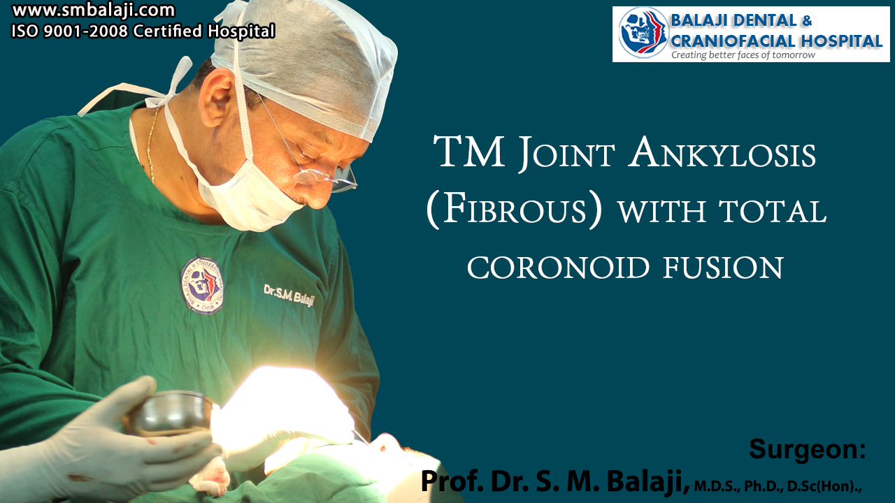
Her 3DCT scan revealed bilateral TMJ fibrous ankylosis between the mandibular condyle and glenoid fossa. The recently inability to move her mandible was due to bilateral coronoid fusion.
Treatment planning formulated and surgical consent obtained from patient
In view of the patient’s extremely limited mouth opening, it was advised that the patient undergo bronchoscopic intubation for anesthesia. This would be followed by releasing the fibrous ankylosis in her bilateral temporomandibular joint along with bilateral coronoidectomy.
This would correct her mouth opening difficulty. Treatment rationale was explained to the patient and her parents in detail and they consented for surgery.
Surgical correction of the bilateral fibrous ankylosis of the TMJ
Bronchoscopic intubation was performed because of the patient’s extremely reduced mouth opening. Following successful induction of general anesthesia, attention was turned to release of the patient’s fibrous ankylosis.
Incisions were first made in the mandibular retromolar region bilaterally. A flap was then elevated and the fibrous ankylosis identified. This was released followed by bilateral coronoidectomy. Passive mouth opening of up to 50 mm was achieved following the surgery. Pterygoid muscle and masseter muscle function was normal.
Successful surgical outcome with increased mouth opening
Hemostasis was achieved and closure done using resorbable sutures. Suction drain was also placed to prevent an accumulation of exudates or blood postoperatively. The patient was then extubated and brought to the recovery room in stable condition.
Total patient satisfaction with the results of the surgery
A mouth opening of about 50 mm was achieved through the surgery. Physiotherapy was started with jaw exercises under supervision after a period of about one week following surgery. Physiotherapy is imperative to prevent relapse of her condition. This would be performed along with mouth opening exercises at subsequent visits.
She and her parents expressed complete understanding of the instructions. They expressed how this surgery would completely transform her life. She would now be able to participate in all social interactions and lead a completely normal life.
A 23-year-old girl from Guntakal with facial deformity
The patient is a 23-year-old female from Guntakal in Telangana, India. She had fallen down and injured her jaw while playing with friends as a little girl. The patient had landed on her chin and had cried incessantly for a few days.
Her parents had taken her to a nearby hospital where they had prescribed her analgesics. She soon returned to her normal playful ways once the patient had subsided.
As the patient grew up, her parents began noticing that her lower jaw growth was lagging. It soon reached the point where she had gross deformity of her lower jaw. Her mouth opening was also very much reduced and she began to have difficulty eating and with speech. There was rigidity to the position of her lower jaw.
TMJ ankylosis surgery of her jaw joint
Her parents had taken her to an oral surgeon in Hyderabad for consultation. He had diagnosed the patient to have left-sided TMJ ankylosis. There was a deviation of her mandible to the left side. He had recommended surgical release of her ankylosed joint. Her parents had consented to surgery and she had undergone TMJ ankylosis surgery.
She now had a degree of facial asymmetry that interferes with her activities of daily living. She has lost all self confidence and rarely leaves the house. This had worried her parents endlessly and they had presented to a local oral surgeon for consultation regarding her problem.
Upon examining the patient, he had realized the degree of correction needed for the patient was extreme. Explaining this to the patient, he had explained the mechanism of distraction osteogenesis surgery to them. He had then referred them to our hospital for facial asymmetry surgery to correct her facial deformity.
Our hospital is a specialist center for facial deformity correction in India. Hundreds of people with facial deformity are rehabilitated in our hospital through distraction osteogenesis and Le Fort osteotomy surgery. Many patient have provided testimony about how surgery at our hospital has completely transformed their lives.
Initial presentation and examination at our hospital
Dr SM Balaji, facial deformity surgeon, met with the patient and her parents and obtained a detailed history from them. This was followed by a thorough clinical examination of the patient’s lower face. He then ordered comprehensive imaging studies for the patient including a 3D CT scan.
The patient was diagnosed with a mandibular asymmetry caused by the TMJ ankylosis. This had caused a shift of the mandible to the left side. There was also an obvious facial asymmetry as a result of this. The patient had minimal mouth opening with poor oral hygiene.
There was also an obvious occlusal cant that had resulted from the long standing ankylosis. Imaging studies revealed that the right side of the mandible was longer than the left side.
Treatment planning explained in detail to the patient
The patient would need treatment on the left side even though the defect was on the right side. This correction would be achieved through mandibular distraction on the left side. A mandibular ramus distractor would be fitted on the left side of the mandible. The patient and her parents expressed understanding of the treatment and consented to surgery.
Successful correction of the facial asymmetry through distraction
Under general anesthesia, an incision was made in the left mandibular retromolar region with elevation of a mucoperiosteal flap. Bone cuts were then made to the outer cortex of the ramus of the mandible. This was followed by fixation of the mandibular ramus distractor using titanium screws.
The inner cortex of the mandible was then cut to facilitate movement of the proximal bone segment. Extreme care was taken throughout to prevent any injury to the inferior alveolar nerve.
A sulcular incision was then made in the maxilla. This was followed by Le Fort I osteotomy. The maxilla was then mobilized and the posterior end of the left maxilla was fixed using transosseous wires. Hemostasis was achieved and closure was done.
Postsurgical treatment phase for the patient
Interarch wiring was done to stabilize the maxillary segment. A latency period of about six to seven days was then given. Following completion of the latency period, the distractor was activated by 1 mm every day.
After achieving the desired increase in length of about 23 mm, the distraction process was stopped. Two weeks after this, a straight plate was fixed to the left posterior maxilla to prevent further downward movement.
There was complete correction of the occlusal cant at the end of this procedure. Radiographs were then obtained after a period of about four months. This revealed complete consolidation of bone at the distraction site in the mandible.
Radiographic evaluation revealed complete bone formation with a patent nerve canal. It was then decided to remove the distractor.
Removal of distractor after bony consolidation
Under general anesthesia, an incision was made followed by elevation of a flap. The distractor was then exposed and successfully removed without incident. The flap was then closed using sutures and the patient recovered from general anesthesia without incident.
Return of self confidence and confidence following successful surgery
The patient and her parents were very happy with the outcome of the surgery. She now had a very pleasant face with symmetrical profile. Her self confidence levels had improved dramatically within a short period of time. She said that she could now face the world bravely and expressed her thankfulness for restoring normalcy back to her life.
The patient is a now 13-year-old boy from Gudur in Telangana, India who had fallen down on his chin as a child. His parents had noticed the development of a facial asymmetry in their son ever since the fall. This fall had been followed by a period of pain and difficulty opening his mouth. However the TMJ pain and TMJ symptoms gradually improved with time resulting in the parents neglecting to seek medical attention for this fall. The facial asymmetry has been gradually worsening and it had now become esthetically unappealing. The parents decided to seek medical help as this was leading to a lot of bullying at school.
It is very important for parents to note that complicated surgical management of TMJ ankylosis can be avoided if optimal medical treatment is given at the time of the initial injury to the TMJ. Whenever a child falls on his chin and complains of even minor pain in the jaw joint, it would be prudent to take the child to the hospital for a checkup. Treatment initiated at this time will help avoid the development of complications later.
Parents approach a local oral surgeon for correction of the facial asymmetry
They had approached a local oral surgeon when he was around 4 years old for correction of the deformity. He had examined the patient and explained to the parents that the patient had ankylosis of the right temporomandibular joint. The oral surgeon had further advised the patient’s parents that he needed to get this addressed at a specialty temporomandibular joint surgery hospital for optimal surgical management. He had thus referred them to Balaji Dental and Craniofacial Hospital for further management.
A gap arthroplasty surgery using a temporalis muscle flap interpositioning had been performed by Dr SM Balaji with complete resolution of the patient’s ankylosis. The patient had been advised to return at a later date for condylar reconstruction surgery using costochondral rib graft. This would enable growth of the patient’s condyle. The patient now returns to our hospital for this surgery.
Injury to the TMJ always leads to growth discrepancies that affect the mandibular condyle and mandibular body. When growth of the mandible is affected, mandibular distraction osteogenesis is performed to set right the discrepancy and reestablish normal facial form and symmetry. When the ankylosis is bilateral, it can result in extreme micrognathia leading to even partial airway obstruction.
Internal devices are always preferred over external devices because of patient convenience. A consolidation phase is always given after fixation of distraction devices to ensure stabilization of bone structure following surgery. This falls under the purview of oral and maxillofacial surgery. Distraction is initiated after completion of the latency period.
Patient presents for initial consultation and treatment planning at our hospital
Dr.SM Balaji, distraction osteogenesis surgery specialist, examined the patient and ordered radiological studies. He explained to them that the child needed to undergo ramus distraction osteogenesis for correction of the facial asymmetry following the condylar reconstruction surgery. This would help induce growth of the right condyle. Therefore, a costochondral rib graft would need to be harvested to create a condyle and to reinforce the mandibular ramus. This would create a primary growth center to induce the growth of the mandible. There was also the presence of a hyperplastic right coronoid process.
Surgical reconstruction of the condyle and ramus
Under general anesthesia, a right inframammary incision was made and a costochondral rib graft harvested. A Valsalva maneuver was next performed to ensure that there was no perforation into the thoracic cavity following which closure was done in layers. This was followed by a right submandibular incision through which dissection was done up to the condyle. The hyperplastic coronoid was reduced and a coronoidectomy was done. The condyle was reconstructed using the rib graft and fixed using titanium screws. The ramus of the mandible was also augmented with a rib graft. Closure was then done using resorbable sutures.
Successful outcome of the surgical procedure
The surgery was a complete success. The condyle was successfully created and ramus augmented. Postoperative OPG taken 2 months after the surgery revealed sufficient bone consolidation. The patient also showed a more symmetrical face following grafting. Distraction osteogenesis to obtain complete facial symmetry will be performed after a period of about 6 months.
Falls are common in childhood. However, a child’s bones are more flexible than adult bones and thus the chances of fractures are lesser than adults. Adult bones are denser and more mineralized that the bones of children.
However, there are certain injuries that are more common in children, which lead to complications. Injuries to the chin where there is direct impact from a fall can lead to injuries to the TMJ. When the impact is sufficiently high, this can lead to bleeding into the TMJ. In many instances, the child might not complain too much of pain in the TMJ. The parents might not be aware of the seriousness of the injury because of this and do not seek medical attention.
Consequences of injury manifesting with passage of time
It is only the passage of time that reveals the serious consequences that arise from this injury. The bleeding into the joint space results in bony fusion of the joints with the glenoid fossa leading to arrest in the growth of the lower jaw and difficulties with eating and speech.
Most cases of bony ankylosis of the mandible that arises in childhood could have been averted with timely medical help. It is therefore very important to seek medical help if such an injury occurs even if the child does not exhibit pain or too much discomfort in the period immediately after the fall.
The consequences of neglecting such an injury are high with the child needing to undergo multiple surgeries along with the psychosocial stress that arises from having facial deformity because of this.
Initial diagnosis of bilateral TMJ ankylosis and sleep apnea
This is a 7-year-old boy from Meghalaya in India who had first presented to Balaji Dental and Craniofacial Hospital in Chennai at the age of 5 with the chief complaint of a retruded mandible and displacement of the tongue to the back of the mouth. This boy had fallen down on his chin as an infant. This had been neglected by his parents at that time. Over time, his jaw growth stopped and he was unable to open his mouth to any degree. They had taken him to a facial plastic surgeon in his hometown. A plastic surgeon in India normally performs a wide variety of surgeries. He however informed them that he performed only cosmetic surgery and referred them here as our hospital is renowned for jaw correction surgery in India. Our hospital rigorously follows the protocols laid down by the American Association of Oral and Maxillofacial Surgeons.
Dr SM Balaji, jaw reconstruction surgeon, examined the patient and obtained a detailed history from the parents. His parents said that the boy periodically stopped breathing during sleep and resumed his breathing with a loud gasp. The patient was diagnosed with bilateral TMJ ankylosis and sleep apnea resulting from backward placement of the tongue due to the micrognathia.
Need for multistage surgical correction explained to the patient
Sleep apnea is a potentially serious sleep disorder in which breathing repeatedly stops and restarts. It was explained to them that surgery needed to be done in two steps for complete correction of the patient’s problems. Orthognathic surgery or sleep apnea correction surgery was performed first as it had to be corrected first. Release of the bilateral TMJ ankylosis would be performed at a later date. A tracheostomy had to be performed due to his restricted mouth opening following which bilateral mandibular body distraction osteogenesis surgery corrected his micrognathia. Bone grafts would not be needed taking into account the age of the patient.
Parents present with patient for TMJ ankylosis surgery
The patient presented with his parents now for corrective jaw surgery for release of his bilaterally ankylosed TMJ. He still has difficulty eating as he was still unable to open his mouth. His parents stated that he has been on a predominantly liquid diet and has lost quite a bit of weight because of this.
Presurgical diagnostics and treatment plan for the patient
Dr SM Balaji examined the patient and ordered comprehensive imaging studies. The patient’s 3D CT revealed that there was complete bony ankylosis between the mandibular condyle and glenoid fossa. His mouth opening was zero due to bilateral TMJ ankylosis. It was decided to perform a tracheostomy as intubation would not be possible due to nil mouth opening. TMJ ankylosis surgery would be performed following the tracheostomy.
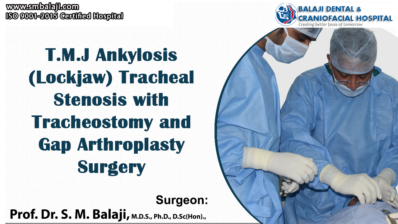
Surgical release of the bilateral TMJ ankylosis
Under general anesthesia, a horizontal skin incision was first made through the previous tracheostomy scar. Minimal dissection was carried out and the strap muscles were retracted. A tracheal incision was made and maturation sutures placed. A tracheostomy tube was then inserted following which skin closure was done in layers.
Attention was then turned to the bilaterally ankylosed temporomandibular joints. An incision was first made in the submandibular region bilaterally and dissection was performed down to the temporomandibular joint. The ankylosed bone was osteotomized bilaterally and gap arthroplasty was performed. Following excision of the bony mass, mouth opening was achieved passively to about 35-40 mm. The incisions were then closed using sutures.
Postsurgical care for the patient
Mouth opening of about 3.5 mm was achieved at the time of surgery. Physiotherapy was initiated for the patient after surgery with jaw exercises under supervision. Mouth opening exercises were also performed for about a month with regular follow up.
The parents were very happy and satisfied with the results. They expressed their delight that he was able to eat normal food without any restrictions now and has slowly begun to gain weight since then. They were instructed to return after six months for a final checkup at the hospital