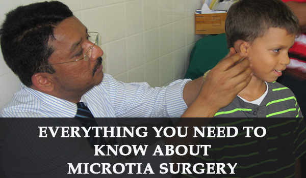
Everything You Need to know about Microtia Surgery

Everything You Need to know about Microtia Surgery

Centre for Excellence in Microtia Surgery in India

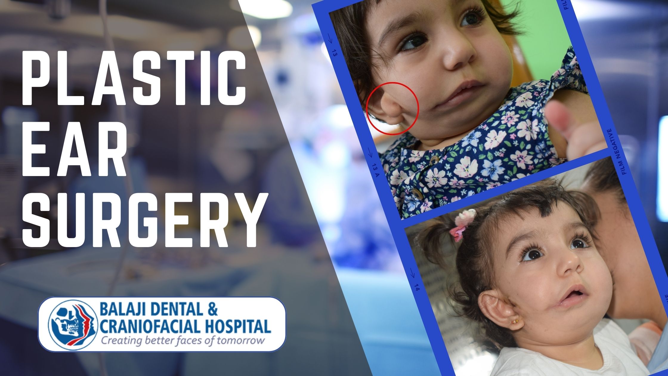
Congenital underdevelopment of the external ear is known as microtia. It is seen in approximately 1 out of 10000 live births around the world. This condition can either be unilateral or bilateral. The right side is more commonly affected when it is unilateral.

It is known as anotia when there is a complete absence of the external ear. Surgical reconstruction of anotia also follows the same principles that are used for microtia repair.

Microtia can vary in severity ranging from grade I-IV. Grade I microtia manifests as a very mild deformity. Grade IV microtia exhibits a complete absence of the external ear and ear canal. The cost of microtia surgery in India varies with the severity of the deformity.

Microtia surgery in India is offered by only a few superspecialty hospitals. The cost of microtia surgery in India is very nominal when compared to the developed Western countries. Microtia correction involves surgical reconstruction of the deformed external ear through ear reconstruction surgery.


Our hospital is a leading facial plastic surgery hospital in India where microtia reconstruction is performed. We have been performing microtia surgery for over 30+ years now. This is one of the specialty cosmetic procedures performed in our hospital.

We have rehabilitated scores of children and adolescents with microtia deformity. Recognized as a premier center for microtia surgery in India, patients from all over the world are referred to our hospital. We have been awarded as a leading facial plastic surgery hospital by many international associations

Possible Etiology behind the Occurrence of Microtia Deformity

Microtia can occur due to genetic defects in either a single gene or multiple genes. There is also a risk of microtia in women with gestational diabetes. It can also be found in conditions like hemifacial microsomia. A mild grade I microtia is difficult to diagnose at the time of birth.

Hearing is also affected in most cases of severe microsomia. Conductive hearing loss is seen in grade II microtia due to stenosis of the external ear canal. It is most severe in grade IV microtia with a complete absence of the external ear canal and the eardrum. Cochlear implants are used to restore hearing for children who are afflicted with the more severe forms of microtia.

Centre of Excellence for Microtia Surgery in India

Situated in Chennai, India, we are a leading center for facial plastic surgery in India. We specialize in facial reconstructive surgery as well as facial cosmetic surgery. Our center is a tertiary referral center for the correction of facial deformities.

Organizations such as the International Cleft Lip and Palate Foundation of Japan and the Dallas-based World Craniofacial Foundation of USA refer patients to us.

This is in lieu of the excellent long-term results for patients undergoing surgeries like ear reconstruction surgery in our hospital. The cost of microtia surgery in India is another factor that makes our hospital the first choice for many international patients.

Our hospital utilizes facial implants for a variety of cosmetic procedures. However, only autologous grafts harvested from the patient are used for microtia surgery. This gives the best results than the utilization of prosthetic ears.

Our hospital is a leading center for the correction of ear deformities. Optimal esthetic results are obtained to ensure total patient satisfaction. Bat ear correction is also performed in our hospital with very good cosmetic results.

Dr. SM Balaji, Ear Reconstruction Surgeon, has over 30+ years of rehabilitating children with congenital defects of the facial region. Our hospital is a renowned center for microtia surgery in India.

Microtia Surgery Utilizing the Two-Stage Technique for Optimal Results

We utilize the two-stage repair for microtia surgery in our hospital. This is a specialized surgical technique for ear reconstruction that is offered only by a few centers offering microtia surgery in India. Surgeons have to undergo many years of training before gaining mastery over this technique.

The first stage involves the harvesting of a costal cartilaginous graft. A template is then prepared using the normal ear as a guide in the case of unilateral microtia.

Construction of symmetrical templates with the creation of cartilaginous structures to recreate the ear is performed for patients with bilateral microtia. The templates help create symmetry in both outer ears through careful crafting of the rib cartilages.

The grafts are then crafted to replicate the structure of the normal ear. This is then inserted into a skin pocket that is created at the site of the deformed ear.

A drain is placed to create negative pressure to prevent the creation of dead space. The shape of the ear framework will be defined without redundancy. Excessive cartilage is banked to be utilized in the second stage repair.

The second stage of surgery is performed in four months’ time. The banked cartilage is first removed under general anesthesia. The cartilaginous structure that was placed in the skin pocket is then raised. The next step entails the utilization of a full-thickness skin graft to recreate the tissue behind the raised ear.

This results in the perfect recreation of asymmetrical ear structure at the site of the deformed ear. The cosmetic transformation is dramatic for the patient.

Leading Center for Facial Plastic Surgery in India

Microtia deformity is often debilitating to the patient. Patients with this deformity, especially children, can face difficulties in social settings as a result of nonacceptance by their peers. This could lead to withdrawal and depression with long-term repercussions.

Our hospital is also a leading research center for facial cosmetic surgery in India. We are considered to be the premier center for microtia surgery in India. Many surgical innovations have evolved in microtia surgery from the research undertaken at our hospital. These innovations have been adopted as the standard operating procedures by many surgeons around the world.

Development of a Dedicated Team for Microtia Surgery in India

Ably led by Dr. SM Balaji, facial plastic surgeon, our surgical team consists of dedicated medical and paramedical staff who have over 30+ years dealing with pediatric patients. Our pediatric anesthesiologists have extensive experience in dealing with children.

Our commitment to providing the best outcomes for our patients is unmatched. We pride ourselves on our dedication and empathy towards all our patients. Utmost importance is given to complete patient satisfaction at our hospital.
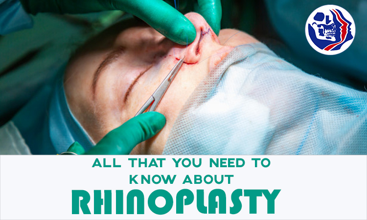
All That You Need to Know About Rhinoplasty

All That You Need to Know About Rhinoplasty

What is Rhinoplasty and what are the Indications for Rhinoplasty?

Rhinoplasty is a surgical procedure that is performed to alter the shape of the nose. It is a nose reshaping surgery. This has been performed since time immemorial and is the most common facial cosmetic surgery performed in the world today.

Also referred to as a nose job, this essentially changes the appearance of the nose and improves the overall esthetic appeal of the face. This can be purely cosmetic for the correction of minor deformities. It can also be a life-altering surgery for patients who have nasal deformities arising from cleft lip and palate deformity or trauma to the nasal region.

The popularity of rhinoplasty (nose surgery) has soared over the last 25-30 years with the advent of liberalization of the economy. Improved facial aesthetics leads to greater self-confidence and higher self-esteem for the individual. This automatically leads to a better overall quality of life.

The Development of India as a Preferred Destination for Medical Tourism

India has become a destination of choice for many from around the world due to its high quality of medical care. Many patients flock to the country for cosmetic surgery. In particular, there is a high demand for experienced facial cosmetic surgeons. Another major factor is the economical rhinoplasty cost in India.

Rhinoplasty is one such procedure for which many patients come to our country. This surgery involves alteration of both bone and cartilage. The best rhinoplasty surgeon in India is someone who offers very good cosmetic and functional results for the patient allied with world-class infrastructure and overall level of care. Many surgeons in India perform primary rhinoplasty.

Our hospital is considered to be a center of excellence for rhinoplasty in the country today due to its consistent track record over the years with complete patient satisfaction. Patients refer us to others stating that we are the best hospital for a nose job in India. We are a superspecialty center for the correction of nasal deformities arising from cleft lip and palate deformity.

Best Rhinoplasty Surgeon in India | Surgical Excellence in the Correction of Nasal Deformities

Dr. SM Balaji, Director and Consultant, Balaji Dental and Craniofacial Hospital, Chennai, is considered to be the best rhinoplasty surgeon in India today. This is based on treatment results, patient satisfaction and overall quality of care offered at our hospital in India. He has over three decades of experience in the correction of nasal deformities. Some of the surgical innovations introduced by him have led to improved results and quicker recovery for patients.

Having performed over 25000 rhinoplasty procedures in his career, he is able to intuitively design the nose that best suits each individual patient. The results obtained over the years are a testament to his dexterity and skill. Our hospital is a destination of choice for many international patients who wish to undergo rhinoplasty.

Many patients with nasal deformity not only have compromised esthetics but also compromised function of the nose. The experience of the surgeon is a vital factor in determining the successful outcome of the surgery. Plastic surgeons specialize in this surgery.

Our hospital is a tertiary care center for redo rhinoplasty in India for patients who have had less than optimal results at other surgical centers. Patients with cleft lip and palate deformity who have had compromised results at other treatment centers are offered world-class care at our hospital.

Cosmetic Rhinoplasty for Correction of Nasal Deformities

There are two main surgical techniques for performing rhinoplasty. They are open rhinoplasty and closed rhinoplasty. All incisions are made inside the nose for closed rhinoplasty thus ensuring that there is no visible scarring at the surgical site. Experienced surgeons are able to perform even complex corrective surgery through closed rhinoplasty.

Open rhinoplasty is performed for the correction of deformities with a very complex presentation. This allows greater access to the surgical site and the surgeon is able to directly visualize the structures.

It takes vast experience to be called the best rhinoplasty surgeon in India. With over three decades of experience operating on every possible variety of nasal deformities, our hospital has become synonymous with excellence in rhinoplasty.

Patient Reasons to Opt for Rhinoplasty

Patients can desire to undergo rhinoplasty for a wide variety of reasons. They can range from correction of deformities arising from congenital conditions like cleft lip and palate deformity, nasal deformities caused by trauma or simply cosmetic correction to get the ideal nasal structure.

Classification of Common Nasal Deformities requiring Cosmetic Nasal Surgery

There are a wide variety of nasal deformities. They include a flat nose with a flattened bridge and an extremely wide nose with large nostrils. It could also present as a short nose or an elongated nose. There could be deformities of the tip of the nose like in a parrot beak nose or a crooked nose deformity amongst many others.

Cartilage grafts harvested from the patient are used for the correction of a wide variety of nasal deformities. These grafts are used to augment the bridge of the nose to correct a flat nose, straighten the dorsum of a crooked nose or correct an asymmetrical nose. The nasal cartilages are also trimmed to provide the best possible cosmetic results from the surgery.

Surgery for Broad Nose Correction and Long Nose Correction

Patients who present for this surgery often have nostrils that are excessively wide and reduce the cosmetic appeal of the face. There is also a flattened appearance to the nose due to this deformity. This is corrected by augmentation of the nasal bridge with a graft followed by reduction of nasal width through excision of excess tissue. Longnose correction is obtained by excision of the nasal cartilages to create a nose that is in harmony with the patient’s face.

Correction of Nasal Hump Deformity and Crooked Nose Deformity

Some patients present with a dorsal nasal hump, which reduces the attractiveness of the nose. There is a hump in the nasal bone in these patients. Surgery is performed through the reduction of this nasal hump to straighten the dorsum of the nose. There is a dramatic improvement in appearance following this surgery.

Crooked nose deformity is most often congenital or caused by trauma to the nose. Patients with this deformity are often self-conscious about their appearance and often have breathing difficulties caused by a deviation from the nasal septum. Correction of this deformity often results in a dramatic improvement to their overall quality of life.

Parrot Beak Deformity Correction for Enhanced Facial Esthetics

A parrot beak deformity or Polly beak nose is often characterized by a dorsal hump and a low tip. Its structure resembles that of the upper beak of a parrot. This is mainly a cosmetic procedure. They state that they wish to undergo plastic surgery at the time of presentation. Patients with this nasal appearance are often dissatisfied with their nasal appearance for a long time before presented for corrective surgery.

Though this can be a complicated surgery, the best rhinoplasty surgeon in India who is often vastly experienced in performing such surgeries performs closed rhinoplasty procedures for optimal esthetic results for the patient.


Correction of Nasal Deformities arising from Cleft Lip and Palate

Our hospital is a superspecialty center for cleft rhinoplasty with thousands of successful surgeries having been performed over the years. We are recognized as one of the premier surgical centers for this procedure in the world and Dr. SM Balaji is unequivocally the best rhinoplasty surgeon in India for this procedure.

The Best Overall Surgical Center for Rhinoplasty Surgery in India

We pride ourselves in providing total patient satisfaction for those who undergo rhinoplasty at our hospital. Our surgeon who is ably assisted by a highly experienced team of medical and paramedical staff ensures that the results are esthetically pleasing and functionally optimal for the patient.


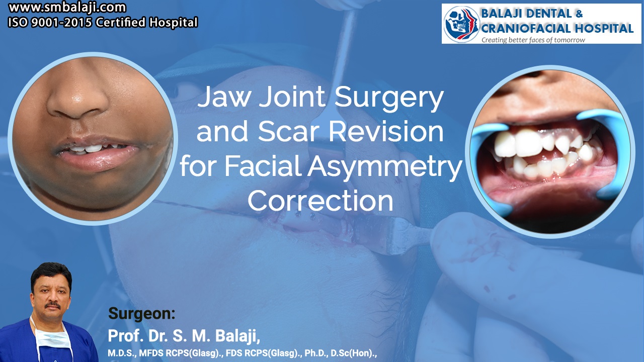
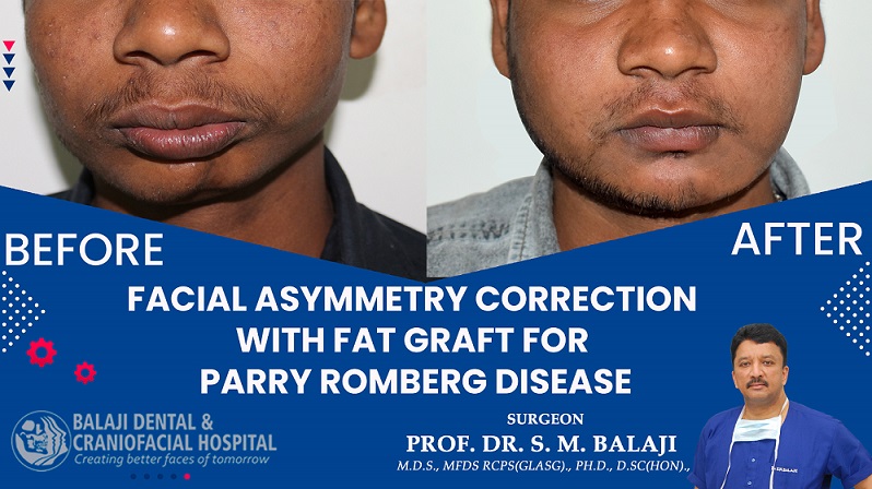
Facial Asymmetry Correction with Fat Graft for Parry Romberg Disease

Facial Asymmetry Correction with Fat Graft for Parry Romberg Disease

What is Facial Asymmetry?

When you examine your face in photos or the mirror, you can find that your features are not perfectly aligned. One ear may begin higher than the other, or one side of your nose may have a sharper point.

Asymmetry refers to having features that do not exactly mirror one another on both sides of the face.

Almost everybody has a degree of facial asymmetry. However, some instances of asymmetry are more obvious than others. Asymmetry may be caused by injury, aging, smoking, and other causes. Mild asymmetry that has always existed is considered natural.

However, recent, visible asymmetry can indicate the presence of a serious condition such as Bell’s palsy or a stroke. Continue reading to learn more about the causes of an asymmetrical face, as well as testing and treatment options.

What is Parry–Romberg syndrome?

Parry–Romberg syndrome (PRS) is a rare disorder that causes progressive shrinkage and degeneration of the tissues beneath the skin, typically on one side of the face (hemifacial atrophy), but also affecting other parts of the body.

Facial Asymmetry Correction with Fat Graft for Parry Romberg Disease

This patient first noticed a developing asymmetry due to gradual atrophy of his left lower jaw a few years back and had been diagnosed with Parry Romberg disease. Parry Romberg disease could have a varied etiology including viral, traumatic, nervous system abnormalities or as an autoimmune disease.

He presented to our hospital seeking correction of his facial asymmetry. He was diagnosed to have atrophy of subdermal fat in the left mental/submental region. A full-thickness dermal fat graft was harvested from his inguinal region. This was then inserted into a submental pocket created at the region of the atrophy.

Minimal overcorrection was performed to allow for slight resorption of the fat graft. He was very pleased with the results of the surgery.


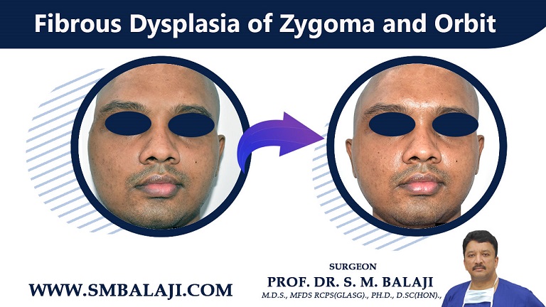
Fibrous Dysplasia of Zygoma and Orbit

Fibrous Dysplasia of Zygoma and Orbit

Young Man Notices Gradual Development of Facial Asymmetry

The patient is a 26-year-old man from Thalassery in Kerala, India. He has had a slowly progressing facial asymmetry for many years now. There was a gradually developing swelling of the right midfacial region.

A biopsy had been obtained and he had been diagnosed with fibrous dysplasia. He had chosen to ignore it as it was not causing him any functional difficulties.

Disfigurement caused by Fibrous Dysplasia and its Probable Etiology

Fibrous dysplasia is a rare disorder of the bone where normal bone is replaced by fibrous tissue. There is an overgrowth of this fibrous tissue, which results in progressive facial disfigurement. The bone is also weakened by this fibrous tissue.

It is thought to result from a genetic mutation at birth but is not hereditary. There is a dull ache in the region of the affected bone. Fractures are also common when it affects the limb bones. The only treatment available is the surgical removal of the fibrous tissue.

Patient Decides to Undergo Surgical Correction of his Deformity

The growth has now progressed to the point where it is causing self-esteem problems for the patient. He was beginning to avoid social interactions because of the extreme facial disfigurement. His parents researched the internet for the best hospital to get this addressed. They also made widespread inquiries regarding the same.

Our hospital is a premiere center for facial plastic surgery in India. Many patients have undergone facial cosmetic surgery and facial reconstructive surgery at our hospital. He and his parents zeroed in on our hospital for his fibrous dysplasia surgery.

Our hospital stringently follows protocols laid down by the American Society of Plastic Surgeons. We also conform to the guidelines laid down by the European Society of Aesthetic Plastic Surgery. Many referrals are made from centers in the European Union to our hospital for correction of facial deformities.

Patients from around the world come to us for reconstructive plastic surgery and facial asymmetry surgery. We utilize advanced facial biometric systems for facial asymmetry correction. Facial implants can be utilized when there is a bony deficiency causing facial asymmetry.

Initial Consultation and Treatment Planning at our Hospital

Dr. SM Balaji, facial cosmetic surgery, examined the patient and spoke with him at length. Biopsies and imaging studies were ordered including CT scans. This revealed a diagnosis of fibrous dysplasia of zygoma and orbit along with the involvement of the temporal bone.

The fibrous overgrowth had caused gross disfigurement of both bones. Our hospital also offers comprehensive imaging services including magnetic resonance imaging (MRI) scan and bone scan.

He explained the surgical treatment process and the cosmetic improvement to the patient. All queries raised by the patient and his family were addressed. The patient and his parents expressed understanding and consented to surgery.

A 3D stereolithographic model of the patient’s skull was obtained. The fibrous overgrowth was studied and presurgical analyses were performed. It was then decided to proceed with surgery.

Surgical Removal of Fibrous Overgrowth

The affected regions of the zygoma and temporal bone were approached through a hemicoronal incision. No other approach would enable satisfactory removal of the fibrous tissue. Surgery progressed through dissection of the galea and temporal fascia. Care was taken to also preserve blood supply to the temporalis muscle to prevent any postsurgical atrophy.

The frontalis and orbicularis oculi branches of the facial nerve were protected throughout the surgical procedure. Dysplastic fibrous overgrowth was removed meticulously. Normal bony contour was established through the removal of the excess tissue. This would restore symmetry to his face.

The fascia was sutured with retraction of the temporal muscle after adequate removal. This was to ensure that there would not be a hollowed-out appearance to the region. The incision was finally closed with staples.

Postsurgical Improvement in Facial Appearance

The patient and his parents were very happy with the results of the surgery. There was a tremendous improvement in the cosmetic appearance of the face. Improvement in the patient’s self-confidence was apparent shortly after surgery.

He and his parents expressed their total satisfaction with the treatment process at our hospital.

Surgery Video


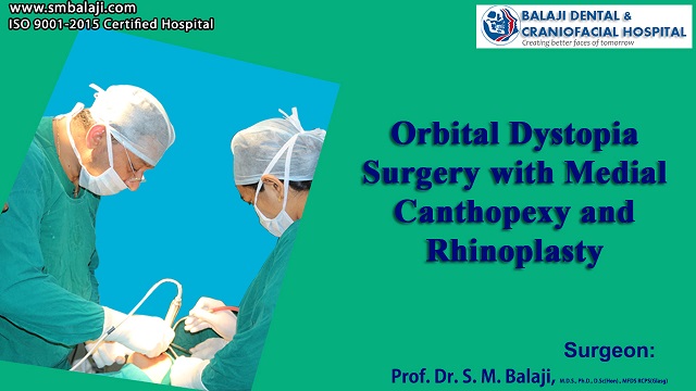
Orbital Dystopia Surgery with Medial Canthopexy and Rhinoplasty

Orbital Dystopia Surgery with Medial Canthopexy and Rhinoplasty

Orbital Dystopia Surgery

One of the most common clinical symptoms of craniofacial malformation is orbital dystopia. The concept dystopia refers to the orbits’ mono- and bilateral asymmetry in at least one of the three-dimensional planes. The clinical examination of the patient is used to make the diagnosis, which is supported by diagnostic instruments such as teleradiography in both normal projections, axial computed tomographic (CT) scans at a rate of 1:1 via the neuro-orbital plan, and three-dimensional CT.

A young man with Grievous Facial Injuries from Road Traffic Accident

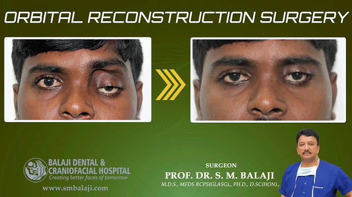
This is a 24-year-old man from Ranchi in Jharkhand, India. He had been involved in a horrific road accident. There was also blindness caused by the loss of his left globe. This had resulted in left-sided facial fractures involving his middle facial region. These fractures involved many bones causing asymmetry.

There was the involvement of his orbital bone, nasal bone, zygoma and maxilla. He had undergone a series of surgeries in various hospitals. These had left him with a facial asymmetry and severe orbital dystopia.

He descended into a depression because of the turn of these events. His worried parents had consulted far and wide for the best hospital to address his problems. They were referred to our hospital by a leading plastic surgeon in their hometown. They contacted our hospital manager and fixed an appointment for a consultation.

Board-certified facial cosmetic surgeons perform these surgeries in Western nations. Oral and Maxillofacial surgeons who are also cosmetic surgeons perform these in India.

Leading Center for Facial Trauma Surgery In India

Our hospital is a leading center for redo facial trauma surgery in India. Many patients with complications arising from surgery performed elsewhere present to our hospital. Our hospital serves as a tertiary referral center for craniofacial surgery in Asia.

We are a specialty center for prosthetic eyeball surgery in India. Our in-house prosthetic laboratory provides custom-made shells to many centers around the country.

Specialty Center for Redo Facial Cosmetic Surgery

Dr SM Balaji, facial cosmetic surgeon, examined the patient and obtained imaging studies. The patient had gross facial asymmetry. This was caused by the malunion of the facial fractures. There was a widening of the canthal bay due to the nasoorbitoethmoidal fracture.

He also had a depressed nose due to a fracture of the nasal bone. CT scans were also obtained for the evaluation of the cribriform plate. Treatment planning was presented to the patient and he consented to surgery.

Facial Asymmetry Correction Performed in our Hospital

A costal cartilage graft was first harvested to augment the depressed nose. The sites of the malunited fractures were then accessed. The plates from the previous surgeries were also removed.

A Titanium mesh with Medpor was then introduced through the subconjunctival incision. This resulted in the reconstruction of the fractured orbital floor. A lateral canthopexy was then performed for the correction of the canthal bay deformity.

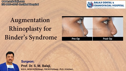
This was next followed by augmentation rhinoplasty. The costal cartilage graft was used to raise the bridge of the flat nose. This resulted in the correction of his nasal deformity. An orbital conformer was then placed at the conclusion of the surgery. This resulted in the correction of his facial deformities.

The patient expressed his satisfaction with the results of the surgery. His parents too conveyed their gratitude before final discharge from the hospital.

Surgery Video


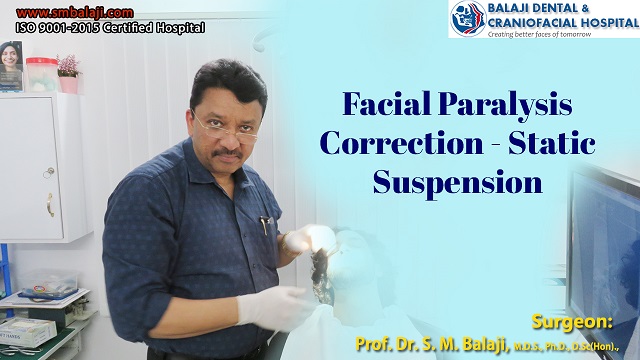
Facial Paralysis Surgery – Static Suspension

Facial Paralysis Surgery – Static Suspension

Facial Paralysis Surgery in India

What is Facial Paralysis?

Bell’s palsy can be caused by a viral infection. It almost never happens more than once.
Bell’s palsy is described by muscle weakness, which causes one-half of the face to droop.
Bell’s palsy normally goes away on its own after six months. Physiotherapy can help prevent muscles from contracting indefinitely.

Patient with Facial Paralysis seeks Solution for his Problem

The patient is a 27-year-old male from Rourkela in Odisha, India. He has had long-standing facial paralysis on the right side of his face. There was a drooping of the corner of the mouth on the affected side. He had tried a variety of medical treatments, which failed to address his problem.

Referral to our Hospital for Treatment of his Condition

This had left him feeling very frustrated. Seeking a solution, he had visited a cosmetic surgeon in his hometown. He underwent a dynamic reanimation with temporalis reanimation surgery. There was no improvement in his facial paralysis and this surgery was a complete failure.

An internet search for the best facial paralysis surgery hospital in India had brought up our name. Having satisfied himself with the search results, he contacted our hospital manager and fixed an appointment for an initial consultation.

Specialty Center for Facial Cosmetic Surgery in India

We are a leading center for facial paralysis surgery in India. Our hospital is well known for facial plastic surgery in India. We are a specialty center for the correction of drooping mouth. This most commonly is caused by facial paralysis.

We offer advanced treatments with the latest innovations from around the world. The cost of lip paralysis surgery in India is a fraction of what it costs in the West. Board-certified plastic surgeons often perform these surgeries in Europe and Japan.

Regaining Movement in Paralyzed Face

Facial reanimation surgery is also performed in our hospital. Patients come from around the world for this surgery. The patient regains the ability to emote following this surgery.

Surgery for Correction of Facial Deformities

Many conditions lead to facial asymmetry. The asymmetry can either be isolated to the soft tissue like in facial paralysis. It can also be due to underlying bony deformities. Facial palsy is caused by loss of innervation to facial muscles on one side supplied by the facial nerve.

Viral infections are a frequent cause of facial palsy (Bell’s palsy), but a majority of such cases recover spontaneously. We also specialize in facial asymmetry surgery. Scores of patients have been rehabilitated after undergoing surgery in our hospital.

Initial Consultation at our Hospital

Dr. SM Balaji, facial cosmetic surgeon, examined the patient. He obtained a detailed history and spoke with the patient at length. Treatment planning was discussed with the patient. The mechanism of static suspension surgery was explained to him.

He was encouraged to ask questions and clear his doubts. Once all his questions were answered to his fullest satisfaction, he expressed understanding and consented to undergo surgery.

Successful Cosmetic Rehabilitation of Facial Paralysis

Surgery commenced with harvesting a fascia lata graft from the thigh. The incision was closed with sutures once the graft was harvested. Attention was next turned to the face. An elliptical incision was made over the nasolabial fold. Orbicularis oris muscle fibers were then identified.

The fascia graft was tunneled up to the zygomatic arch. The graft was then looped around the arch to form a circumzygomatic sling. Care was taken to ensure that there was no damage to any nerves or vessels.

Both ends of the fascia lata sling were then sutured to the orbicularis oris. There was a good improvement of facial symmetry following this procedure.  A lateral tarsorrhaphy was also performed as his cornea was getting ulcerated due to his inability to oppose the left eye.

Total Patient Satisfaction with Results of Surgery

The patient was very happy with the results of the surgery. He expressed his gratitude to the surgical team before discharge from the hospital.

Surgery Video