Oral Submucous Fibrosis, Complete Trismus Release, Excision of Fibrous Bands and Nasolabial Flap Reconstruction
[et_pb_section fb_built=”1″ _builder_version=”3.22″][et_pb_row _builder_version=”3.25″ background_size=”initial” background_position=”top_left” background_repeat=”repeat”][et_pb_column type=”4_4″ _builder_version=”3.25″ custom_padding=”|||” custom_padding__hover=”|||”][et_pb_text _builder_version=”3.27.4″ background_size=”initial” background_position=”top_left” background_repeat=”repeat”]Oral Submucous Fibrosis (OSMF) is a pre-cancerous condition seen predominantly in the Indian subcontinent and South East Asia. In this condition, the deep tissues of the cheeks become thick and fibrosed leading to severely restricted mouth opening, referred to as trismus. ] There is great difficulty in opening the mouth, patients cannot tolerate hot and spicy food, and the cheek lining inside the mouth becomes pale, blanched or marble-like. This is a very serious condition because it has high chances of advancing into mouth cancer (squamous cell carcinoma). The most common cause of this pre-cancerous condition is chewing of tobacco/areca nut. The patient is a middle-aged man with oral submucous fibrosis who presented to Dr. S. M. Balaji, Craniofacial Surgeon, Chennai with complete trismus as a result of which he couldn’t open his mouth more than a few mm. Upon taking a history, it was found that he had been chewing paan or betel quid containing betel leaf, areca nut, and slaked lime for the past 15 years. The patient also complained of a burning sensation in his mouth. Upon palpating his cheeks, thick, tight bands of tissue could be felt lining his cheeks and he had jaw rigidity. Surgical intervention was the only viable option. General anesthesia was given through Flexible Fibreoptic Intubation (FFI) since he had very limited mouth opening. Cuts were placed in the inner cheek and the thickened bands of fibrous tissue were excised. Mouth opening was increased to the normal 3-4 cm using a mouth gag. An inferiorly based nasolabial flap based on the facial artery was taken in such a way that the flap margin fell in the skin fold and post-surgery scar is inconspicuous. The flap was rotated, tunneled into the mouth, and sutured to the inner cheek. The nasolabial incision was closed in layers. This was done on both sides. The patient was prescribed mouth-opening exercises and counseled on complete cessation of the habit. [/et_pb_text][et_pb_video src=”https://www.youtube.com/watch?v=em5jPlYQ1tI” _builder_version=”4.9.4″ _module_preset=”default”][/et_pb_video][/et_pb_column][/et_pb_row][/et_pb_section]
Dentigerous Cyst Enucleation Surgery
[et_pb_section fb_built=”1″ _builder_version=”3.22″][et_pb_row _builder_version=”3.25″ background_size=”initial” background_position=”top_left” background_repeat=”repeat”][et_pb_column type=”4_4″ _builder_version=”3.25″ custom_padding=”|||” custom_padding__hover=”|||”][et_pb_text _builder_version=”3.27.4″ background_size=”initial” background_position=”top_left” background_repeat=”repeat”]This teenage boy had been complaining of pain on the left side of his lower jaw for a few days now. There were a few teeth missing on that side. He was brought to Balaji Dental and Craniofacial Hospital, Teynampet, Chennai, by his parents for treatment. Dr. Balaji examined the patient and ordered diagnostic studies including an OPG, which revealed multiple unerupted impacted teeth and a radiolucent area in relation to the lower left first molar. A 3D axial CT scan was ordered and the diagnosis of a dentigerous cyst was made. It was explained to the patient and his parents that this had to be managed surgically and they were in full agreement with that. The patient was taken to the operating room and general anesthesia was induced. A mucogingival flap was raised and reflected down to the sulcus. The dentigerous cyst was enucleated in its entirely and two unerupted teeth within the cyst were removed. The flap was then sutured and the patient recovered uneventfully from general anesthesia. [/et_pb_text][et_pb_video _builder_version=”4.9.4″ _module_preset=”default” src=”https://www.youtube.com/watch?v=FE0yX-T7utU&t=50s” hover_enabled=”0″ sticky_enabled=”0″][/et_pb_video][/et_pb_column][/et_pb_row][/et_pb_section]
Primary Incomplete Unilateral Cleft Lip and Palate Surgery
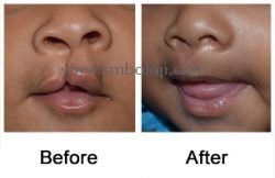
A 3-month old baby girl was brought to our hospital seeking the best possible treatment for incomplete unilateral cleft lip and palate. The parents were greatly worried about their baby girl’s present condition and also complained about the difficulty in feeding her. Cranio-Maxillofacial Surgeon Dr. S.M. Balaji performed the primary incomplete lip repair surgery for unilateral cleft lip using Modified Millard’s technique. Following surgery, the baby appeared as any other normal baby of her age and was able to feed well. The parents were very pleased with the results.
Re-alveolar bone graft Surgery, Fistula Closure and Cleft Rhinoplasty Surgery
[et_pb_section fb_built=”1″ _builder_version=”3.22″][et_pb_row _builder_version=”3.25″ background_size=”initial” background_position=”top_left” background_repeat=”repeat”][et_pb_column type=”4_4″ _builder_version=”3.25″ custom_padding=”|||” custom_padding__hover=”|||”][et_pb_text _builder_version=”3.27.4″ background_size=”initial” background_position=”top_left” background_repeat=”repeat”]This young girl had been born with a left-sided cleft lip, alveolus, and palate. She had undergone repair of her cleft lip as an infant with an alveolar rib graft, but the graft hadn’t fused with the bone and had been a failure. She had developed an asymmetry of her nose because of this and a deficiency in the development of the cartilaginous part of her columella, which had lead to a collapsed left nostril. This had made her very quiet and withdrawn, isolating herself from her peers at school. The alveolar cleft in the region of her left lateral incisor was causing a direct communication with her nasal cavity through an oronasal fistula, which was leading to regurgitation of fluids from her mouth into her nasal cavity. Her parents had been enquiring everywhere as to where her defect would be best set right and had finally been referred to Balaji Dental and Craniofacial Hospital, Teynampet, Chennai. Dr. S. M. Balaji, Cranio-Maxillofacial Surgeon, examined the patient thoroughly and ordered comprehensive imaging studies including a 3D axial CT scan. He then explained the treatment plan in detail to the parents of the patient and they expressed their desire to go ahead with surgery. After satisfactory induction of general anesthesia, two costochondral rib grafts were obtained from the patient. The wound was then closed in layers after ascertaining patency of the pleural cavity through the positive pressure ventilation test. Following this, mucogingival and palatal flaps were raised on the left side of the patient’s maxillary region at the region of the alveolar cleft defect. Costochondral rib grafts were shaped and crafted to fit into the area of bony defect and fixed with screws. Attention was next turned to the collapsed columella. A costochondral rib graft that had been shaped to precisely fit into the columella was inserted along the length of its base and stabilized in place with sutures. This lifted up the collapsed columella of the nose and set right the deformity to the left nostril. The palatal and the mucogingival flaps were then closed with sutures and the patient recovered uneventfully from general anesthesia. The patient expressed her happiness to Dr. Balaji for setting right the deformity to her nose and her parents expressed their gratitude to Dr. Balaji for enabling an improvement in the aesthetic as well as functional quality of life for the patient. [/et_pb_text][et_pb_video src=”https://www.youtube.com/watch?v=ufiBluI_hic” _builder_version=”4.9.4″ _module_preset=”default”][/et_pb_video][/et_pb_column][/et_pb_row][/et_pb_section]
Cleft Rhinoplasty- Nasal Augmentation and Buckling Correction Surgery
[et_pb_section fb_built=”1″ _builder_version=”3.22″][et_pb_row _builder_version=”3.25″ background_size=”initial” background_position=”top_left” background_repeat=”repeat”][et_pb_column type=”4_4″ _builder_version=”3.25″ custom_padding=”|||” custom_padding__hover=”|||”][et_pb_text _builder_version=”3.27.4″ background_size=”initial” background_position=”top_left” background_repeat=”repeat”]The patient is a young girl who was born with a left-sided unilateral cleft lip defect. She was operated upon as an infant at an outside hospital, but that correction had left her with an unsightly residual scar. She had slight lip incompetency on the left side in association with the scar tissue. There was also a buckling of the left nostril. This had lead to taunts and jibes at school from other children, which lead to her becoming withdrawn and quiet. Her worried parents brought her over to Balaji Dental and Craniofacial Hospital, Teynampet, Chennai, where she was thoroughly examined by Dr. S. M. Balaji, Cranio-Maxillofacial Surgeon, who suggested a minor scar revision procedure and nasal augmentation with buckling correction surgery. He explained to them that this involved harvesting a costochondral rib graft from the patient to be used to augment the bridge of the nose and her parents were in full agreement with that. Upon induction of adequate general anesthesia, a costochondral rib graft was harvested and the wound was closed in layers with sutures. Following this, the minor scar revision surgery was performed with release of fibrous bands from the scar tissue, which lead to improvement in the patient’s lip incompetency. Next an intranasal incision was made at the region of the lateral crus and a tunnel was created extending up to the bridge of the nose. The cartilaginous rib graft was then manipulated into position resulting in a straighter profile to the nose along with correction of the nasal buckling of the left nostril. The incision was then closed with sutures. The patient was overjoyed at the results of the surgery and couldn’t stop smiling, thanking Dr. Balaji profusely for the way he had set right her problem. [/et_pb_text][et_pb_video _builder_version=”4.9.4″ _module_preset=”default” src=”https://www.youtube.com/watch?v=Lw6nQ_xlrpk” hover_enabled=”0″ sticky_enabled=”0″][/et_pb_video][/et_pb_column][/et_pb_row][/et_pb_section]
Velopharyngeal insufficiency (Hypernasality) Cleft palate- Pharyngoplasty Surgery(Nasal Speech Correction)
[et_pb_section fb_built=”1″ _builder_version=”3.22″][et_pb_row _builder_version=”3.25″ background_size=”initial” background_position=”top_left” background_repeat=”repeat”][et_pb_column type=”4_4″ _builder_version=”3.25″ custom_padding=”|||” custom_padding__hover=”|||”][et_pb_text _builder_version=”3.27.4″ background_size=”initial” background_position=”top_left” background_repeat=”repeat”]This young man had difficulty pronouncing certain words well and had a nasal twang to his voice. He had been born with a cleft lip and palate and had undergone repair as an infant. He had subsequently developed velopharyngeal incompetence, where there is escape of air through the nose during speech. He presented at Balaji Dental and Craniofacial Hospital, Teynampet, Chennai for correction of his problem. Dr. S. M. Balaji, Cranio-Maxillofacial Surgeon, examined him and explained to him that he needed a pharyngoplasty surgery for correction of his problem. The patient was in agreement with this and was scheduled for surgery. The aim of this surgery is to create a dynamic sphincter in the pharynx by repositioning the palatopharyngeus muscles. The patient was taken to the operating room and underwent general anesthesia without complications. Incisions were made to release the posterior faucial pillars, including the palatopharyngeus muscles, which were then crisscrossed and sutured together on the posterior pharyngeal wall to form a sphincter leaving a small opening or “port” for breathing through the nose. A positive suction test was performed after completion of the surgery and showed new dynamic velopharyngeal sphincter action indicating successful correction of velopharyngeal incompetence. The patient recovered from general anesthesia without complications. [/et_pb_text][et_pb_video _builder_version=”4.9.4″ _module_preset=”default” src=”https://www.youtube.com/watch?v=wFneafecSVU” hover_enabled=”0″ sticky_enabled=”0″][/et_pb_video][/et_pb_column][/et_pb_row][/et_pb_section]
TMJ Ankylosis Gap Arthroplasty Surgery with Coronoidectomy and temporalis interposition Surgery – Sleep Apnea and Snoring
[et_pb_section fb_built=”1″ _builder_version=”3.22″][et_pb_row _builder_version=”3.25″ background_size=”initial” background_position=”top_left” background_repeat=”repeat”][et_pb_column type=”4_4″ _builder_version=”3.25″ custom_padding=”|||” custom_padding__hover=”|||”][et_pb_text _builder_version=”4.9.4″ background_size=”initial” background_position=”top_left” background_repeat=”repeat” hover_enabled=”0″ sticky_enabled=”0″] This young lady was involved in a road traffic accident many years ago in her hometown. First aid had been administered immediately after the accident, but a minor injury to her right TMJ went undiscovered at that time because there were no good diagnostic facilities in her hometown. This resulted in ankylosis of her right TMJ with resultant retarded growth of the mandible on the right side with deviation of the mandible to the right. She developed snoring and sleep apnea over the years and this had now been troubling her for a long time. She had undergone ankylosis release surgery multiple times elsewhere but without much success. Her parents finally brought her to Balaji Dental and Craniofacial Hospital, Teynampet, Chennai, for definitive surgical correction.Dr. S. M. Balaji, Cranio-Maxillofacial Surgeon, examined the patient and ordered diagnostic studies including a 3D axial CT scan. A diagnosis of right-sided ankylosis of the TMJ was made and he explained the surgical procedure in detail to the patient and her parents. They were in complete agreement with the proposed treatment plan and the patient was scheduled for surgery. The patient was taken to the operating theatre. Anesthesia was administered via fiberoptic nasal intubation because of the patient’s inability to open her mouth. This was done to avoid performing a tracheostomy. A submandibular incision was then made just below the margin at the angle of the mandible on the right side to access the TMJ and a gap arthroplasty was performed. The ankylosis was released and a temporalis muscle interpositioning procedure was performed to prevent reankylosis of the joint to the glenoid fossa. Attention was next turned to the left side where the coronoid process was accessed through an intraoral approach. A coronoidectomy was performed to negate the action of the left temporalis muscle and enable increased mouth opening. Full mouth opening was established at the end of the procedure. The incisions were closed with sutures and the patient recovered uneventfully from general anesthesia. The joint was mobilized within a week’s time and the patient was able to slowly begin eating solid foods again. She did not demonstrate any sleep apnea or snoring after the surgery while in the hospital. The patient and her parents expressed their happiness at the results of the surgery before being discharged from the hospital. Surgery Video [/et_pb_text][et_pb_video _builder_version=”4.9.4″ _module_preset=”default” src=”https://www.youtube.com/watch?v=zAecLX5H_g0″ hover_enabled=”0″ sticky_enabled=”0″][/et_pb_video][/et_pb_column][/et_pb_row][/et_pb_section]
Simultaneous Maxillo Mandibular Distractor Removal with Lateral Defect Augmentation With Rib Grafts
[et_pb_section fb_built=”1″ _builder_version=”3.22″][et_pb_row _builder_version=”3.25″ background_size=”initial” background_position=”top_left” background_repeat=”repeat”][et_pb_column type=”4_4″ _builder_version=”3.25″ custom_padding=”|||” custom_padding__hover=”|||”][et_pb_text _builder_version=”4.9.4″ background_size=”initial” background_position=”top_left” background_repeat=”repeat” hover_enabled=”0″ sticky_enabled=”0″] The patient is a young woman with distortion of her lower face due to mandibular asymmetry. This had lead to her socially isolating herself due to people staring at her and passing comments since she was a little girl. She presented to Balaji Dental and Craniofacial Hospital, Teynampet, Chennai, for correction of her condition. Dr. S. M. Balaji, Maxillo-Craniofacial Surgeon, viewed her 3-D axial CT scans and decided to perform simultaneous maxillomandibular distraction for the patient to correct her vertical mandibular deficiency and occlusal cant on the right side. Maxillomandibular distraction was successfully performed and her facial asymmetry was set right, but there was a persistent bone deficiency in the right side of her mandible. She now presents for removal of her distractors as well as for placement of rib grafts for correction of her bone defect. The patient was taken to the operating theatre where general anesthesia was induced. Two rib grafts were taken from the right side for filling in the bony deficiency on the right side of her mandible and the incision was closed in layers with sutures. A right-sided submandibular incision was made and dissection was carried down to the level of the distractor. The distractor was then unscrewed and removed uneventfully. The rib grafts were then shaped and screwed in place to fill in the bone deficiency in the mandible. The patient presented to Dr. Balaji a few months after the surgery and expressed her delight at the way he had given her a new lease of life by setting right her facial asymmetry. Surgery Video [/et_pb_text][et_pb_video _builder_version=”4.9.4″ _module_preset=”default” src=”https://www.youtube.com/watch?v=GOfmILAOem8″ hover_enabled=”0″ sticky_enabled=”0″][/et_pb_video][/et_pb_column][/et_pb_row][/et_pb_section]
Long lower jaw corrected surgically enhancing the facial profile without any scars on the face
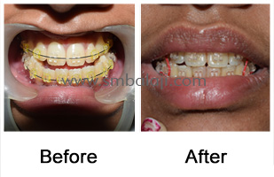
A patient of age 25 years presented to our hospital with complaint of malaligned teeth, forwardly placed lower jaw and inability to close her mouth completely. She also stated that she had speech difficulty along with difficulty in chewing foods. After thorough clinical and radiological examination, Maxillofacial Surgeon Dr. S. M. Balaji planned to correct her bite surgically by removing the mandibular excess. Presurgical orthodontic treatment was started and the teeth were aligned properly. Through intraoral approach, bilateral sagittal split osteotomy (short lingual split technique) was done and the excessive length of the mandible was reduced. This improved her appearance dramatically and she was overjoyed to have her teeth aligned and jaw corrected without any visible scars on the face. Her chewing efficiency and speech also improved drastically.
9 Year old boy from Seychelles gets a new external ear – Ear Reconstruction Surgery
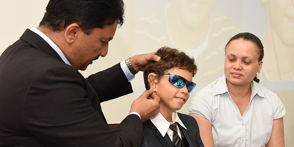
Ear Reconstruction Surgery(Microtia) for a Seychelles boy 9-year-old Nathan Parkash from Seychelles was born with completely absent external portion of his right ear, he only had a very small, rudimentary ear lobule. His left ear was normal. Doctors in his hometown diagnosed that the child’s hearing was normal. As the boy started going to school, his social interactions increased, children at school began teasing him because of his ear. He suffered ridicule and unwelcome stares. He saw that he was not able to wear sunglasses and walkmans like his fellow mates. He started becoming psychologically depressed about his appearance. Slowly he became withdrawn and self-conscious. His parents were becoming increasingly worried for their child’s poor self-esteem. They looked for hospitals that do ear reconstructions all around the world and they found that such centers are very few and the results were not up-to the mark. Usually people from Seychelles go to South Africa or Dubai for medical treatment. They found that very few hospitals were available for doing such procedures but the outcomes were not satisfactory. The Govt. of Seychelles referred them to our hospital for the ear reconstruction surgery (Microtia) as they found the surgical results of the surgeon to be exceptionally best in India. Dr. S.M. Balaji decided to perform complete ear reconstruction in 2 stages using the boy’s own rib graft (costochondral graft) as artificial materials can lead to inherent problems like infection, allergy and loss of stability over time. Using the patient’s own graft is the best technique giving permanent and stable results. The first stage was called cartilage-banking surgery. Based on the measurements of the normal left ear, a template was made. It was important to do this correctly as the ear characteristics have to be accurately replicated to achieve symmetry. A costochondral graft was taken from the rib. Using the template as reference, the graft was contoured to create the structural framework. This is most crucial and has to be done with great precision and accuracy. The skin of the right ear was raised creating a pocket and the cartilage framework was placed below the skin. Additional piece of cartilage was also placed adjacent for future use. A surgical drain was placed that created a suction making the overlying skin to adapt to the framework and the ear shape was achieved After 3 months the second stage surgery called ear-lifting surgery was done. Using the excess preserved cartilage and skin graft, the entire ear framework was lifted from the side of the head giving a normal, raised appearance to the ear. Following reconstruction the ear was given a more aesthetic, natural-looking appearance. As the boy grows the normal left ear will grow, but the reconstructed right ear will not increase in size. So to avoid re-surgery, the right ear was reconstructed with a higher dimension matching the adult size. Following second stage surgery the boy is delighted about his “new ear”. Also he is happy that he won’t be teased any longer. He and his parents are happy with the surgery outcome and returned to their hometown. He may require revision surgery at later stage. Ear reconstruction surgeries are challenging and must be performed with skill, care & artistic sense. Timely correction of ear defects is important for the person’s physical and emotional well-being. Surgery Video:
